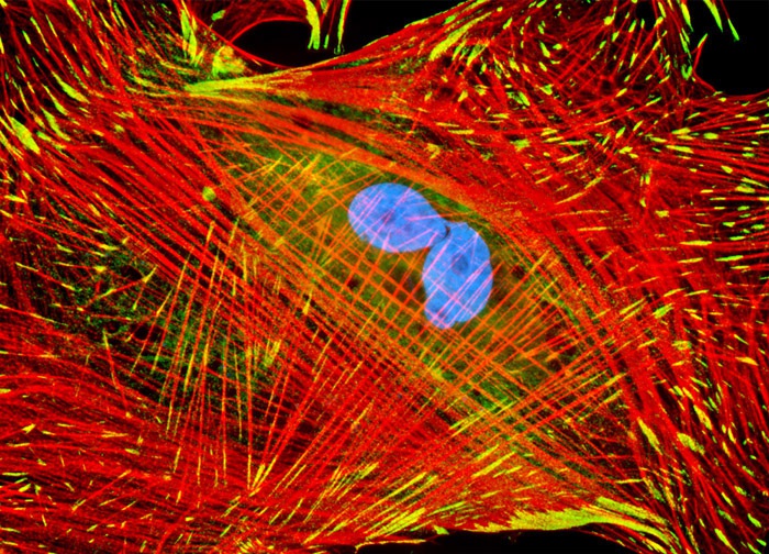
Indian Muntjac Deer Skin Fibroblast Cells
Focal adhesions and adherens junctions were visualized in a culture of Muntjac cells (shown above)immunofluorescently labeled with primary anti-vinculin mouse monoclonal antibodies followed by goat anti-mouse Fab heavy and light chain fragments conjugated to Cy2. In addition, the specimen was stained for DNA with Hoechst 33342 and for the cytoskeletal filamentous actin network with Alexa Fluor 568 conjugated to phalloidin. Images were recorded in grayscale with a 12-bit digital camera coupled to either a Nikon E-600 or Eclipse 80i microscope equipped with bandpass emission fluorescence filter optical blocks. During the processing stage, individual image channels were pseudocolored with RGB values corresponding to each of the fluorophore emission spectral profiles.
Featured in:
Share this page:


