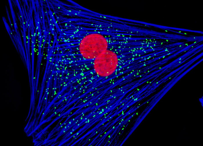
Indian Muntjac Deer Skin Fibroblast Cells
The log phase monolayer culture of Indian Muntjac cells illustrated above was fixed, permeabilized, and blocked with 10-percent normal goat serum in phosphate-buffered saline prior to immunofluorescent labeling with rabbit primary antibodies to PMP 70, a protein resident in peroxisomal membranes, and mouse primary antibodies to tubulin, the basic structural constituent of microtubules. The culture was subsequently stained with a mixture of goat anti-rabbit and anti-mouse secondary antibody fragments (heavy and light chain) conjugated to Oregon Green 488 and Texas Red, respectively. In addition, the culture was labeled for the filamentous actin network with Alexa Fluor 350 conjugated to phalloidin, yielding blue fluorescence emission. Images were recorded in grayscale with a 12-bit digital camera coupled to either a Nikon E-600 or Eclipse 80i microscope equipped with bandpass emission fluorescence filter optical blocks. During the processing stage, individual image channels were pseudocolored with RGB values corresponding to each of the fluorophore emission spectral profiles.
Featured in:
Share this page:


