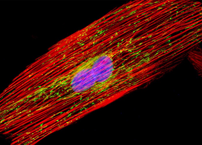
Indian Muntjac Deer Skin Fibroblast Cells
The culture of Indian Muntjac deer skin fibroblasts presented in the digital image above was transfected with a pEYFP-Mitochondria (enhanced yellow fluorescent protein) chimeric plasmid subcellular localization vector. After fixation and permeabilization, the cells were labeled with Alexa Fluor 568 conjugated to phalloidin and DAPI, which target filamentous actin and DNA in the cell nucleus, respectively. Images were recorded in grayscale with a 12-bit digital camera coupled to either a Nikon E-600 or Eclipse 80i microscope equipped with bandpass emission fluorescence filter optical blocks. During the processing stage, individual image channels were pseudocolored with RGB values corresponding to each of the fluorophore emission spectral profiles.
Featured in:
Share this page:


