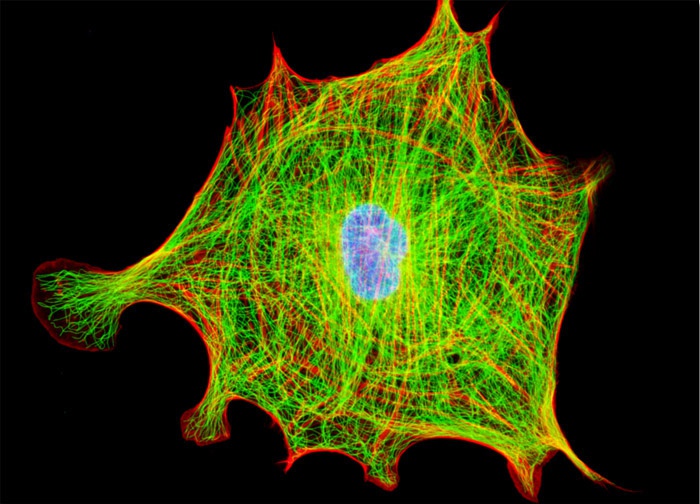
Indian Muntjac Deer Skin Fibroblast Cells
Indian Muntjac cellular tubulin was visualized in the culture of fibroblasts depicted in the digital image above by immunofluorescent labeling with primary anti-tubulin mouse monoclonal antibodies followed by goat anti-mouse secondary antibody fragments conjugated to Alexa Fluor 488. The cell culture was also labeled for the cytoskeletal filamentous actin network with Alexa Fluor 546 conjugated to phalloidin. Cell nuclei were counterstained with TO-PRO-3. Images were recorded in grayscale with a 12-bit digital camera coupled to either a Nikon E-600 or Eclipse 80i microscope equipped with bandpass emission fluorescence filter optical blocks. During the processing stage, individual image channels were pseudocolored with RGB values corresponding to each of the fluorophore emission spectral profiles with the exception of TO-PRO-3, which was pseudocolored cyan.
Featured in:
Share this page:


