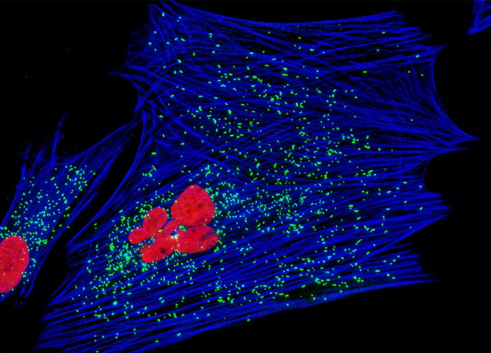
Indian Muntjac Deer Skin Fibroblast Cells
A culture of Indian Muntjac cells (illustrated above) was triple-labeled using double immunofluorescence and a phallotoxin. Microtubules were visualized with mouse anti-tubulin primary antibodies, while peroxisomes were stained with rabbit anti-PMP 70 (peroxisomal membrane protein) antibodies. The secondary antibodies utilized were goat anti-mouse and anti-rabbit conjugated to Texas Red and Oregon Green 488, respectively. The F-actin network was counterstained with Alexa Fluor 350 conjugated to phalloidin. Images were recorded in grayscale with a 12-bit digital camera coupled to either a Nikon E-600 or Eclipse 80i microscope equipped with bandpass emission fluorescence filter optical blocks. During the processing stage, individual image channels were pseudocolored with RGB values corresponding to each of the fluorophore emission spectral profiles.
Featured in:
Share this page:


