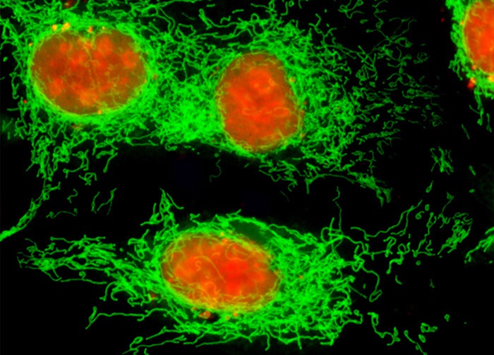
Madin-Darby Canine Kidney Epithelial Cells (MDCK Line)
The culture of Madin-Darby canine kidney epithelial cells appearing in the digital image above was transfected with a pEYFP-Mitochondria chimeric plasmid subcellular localization vector. The cells were also stained with SYTOX Orange, targeting DNA in the cell nucleus. Images were recorded in grayscale with a 12-bit digital camera coupled to either a Nikon E-600 or Eclipse 80i microscope equipped with bandpass emission fluorescence filter optical blocks. During the processing stage, individual image channels were pseudocolored with RGB values corresponding to each of the fluorophore emission spectral profiles.
Featured in:
Share this page:


