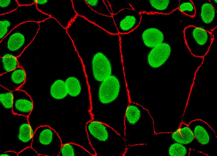
Madin-Darby Canine Kidney Epithelial Cells (MDCK Line)
Simultaneous localization of tight junctions and nuclear pore complex proteins (NPCP) was performed in a double immunofluorescence experiment with the MDCK cell culture illustrated above using mouse anti-NPCP and rabbit anti-ZO-3 primary antibodies. The subcellular targets were visualized using goat anti-mouse and anti-rabbit secondary antibodies (IgG) conjugated to Alexa Fluor 488 and Alexa Fluor 568, respectively. Images were recorded in grayscale with a 12-bit digital camera coupled to either a Nikon E-600 or Eclipse 80i microscope equipped with bandpass emission fluorescence filter optical blocks. During the processing stage, individual image channels were pseudocolored with RGB values corresponding to each of the fluorophore emission spectral profiles.
Featured in:
Share this page:


