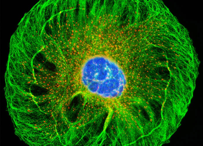
Madin-Darby Canine Kidney Epithelial Cells (MDCK Line)
The adherent log phase culture of MDCK cells illustrated above was treated for one hour with MitoTracker Red CMXRos in order to label the mitochondrial network, and the fixed cells were then incubated with mouse anti-cytokeratin primary antibodies followed by goat anti-mouse secondary antibodies (IgG) conjugated to Cy2. Nuclei were counterstained with Hoechst 33258. Images were recorded in grayscale with a 12-bit digital camera coupled to either a Nikon E-600 or Eclipse 80i microscope equipped with bandpass emission fluorescence filter optical blocks. During the processing stage, individual image channels were pseudocolored with RGB values corresponding to each of the fluorophore emission spectral profiles.
Featured in:
Share this page:


