Madin-Darby Ovine Kidney Epithelial Cells (MDOK Line)
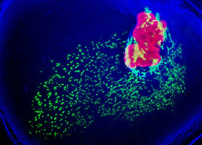
Nuclear histone proteins were targeted in the culture of MDOK cells presented above with mouse anti-histone (pan) monoclonal antibodies, which were imaged with goat anti-mouse Fab fragments conjugated to Texas Red (labeling the nucleus). The specimen was simultaneously labeled for the Golgi complex with Alexa Fluor 488 conjugated to goat secondary antibodies that target rabbit anti-giantin. Alexa Fluor 350 conjugated to phalloidin was utilized to counterstain the cytoskeletal F-actin network. Images were recorded in grayscale with a 12-bit digital camera coupled to either a Nikon E-600 or Eclipse 80i microscope equipped with bandpass emission fluorescence filter optical blocks. During the processing stage, individual image channels were pseudocolored with RGB values corresponding to each of the fluorophore emission spectral profiles.
Featured in:
Share this page:


