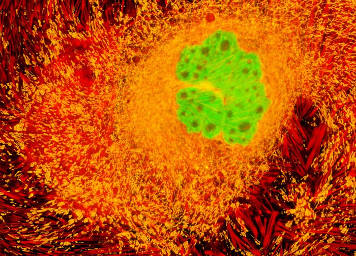
Madin-Darby Ovine Kidney Epithelial Cells (MDOK Line)
The filamentous actin components of the cytoskeletal framework were labeled in a culture of MDOK epithelial cells (shown above) with Alexa Fluor 633 conjugated to phalloidin. Mitochondria and DNA in the cell nucleus were also probed with MitoTracker Red CMXRos and SYTOX Green, respectively. Images were recorded in grayscale with a 12-bit digital camera coupled to either a Nikon E-600 or Eclipse 80i microscope equipped with bandpass emission fluorescence filter optical blocks. During the processing stage, individual image channels were pseudocolored with RGB values corresponding to each of the fluorophore emission spectral profiles.
Featured in:
Share this page:


