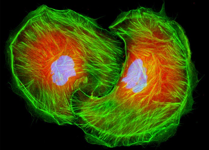
Madin-Darby Ovine Kidney Epithelial Cells (MDOK Line)
A fixed and permeabilized monolayer culture of MDOK cells (illustrated above) was treated with mouse anti-nonmuscle myosin II (heavy chain) monoclonal primary antibodies followed by goat anti-mouse secondary antibodies (IgG) conjugated to Texas Red-X. Oregon Green 488 conjugated to phalloidin was included in the secondary cocktail to target the filamentous actin network. After the immunofluorescence and phallotoxin labeling, the cells were washed and counterstained with Hoechst 33258, targeting DNA in the cell nucleus. Images were recorded in grayscale with a 12-bit digital camera coupled to either a Nikon E-600 or Eclipse 80i microscope equipped with bandpass emission fluorescence filter optical blocks. During the processing stage, individual image channels were pseudocolored with RGB values corresponding to each of the fluorophore emission spectral profiles.
Featured in:
Share this page:


