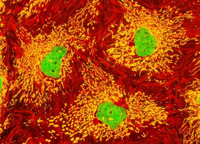
Madin-Darby Ovine Kidney Epithelial Cells (MDOK Line)
Three fluorescent probes were utilized to label the Madin-Darby ovine kidney cell culture depicted in the digital image above, including MitoTracker Red CMXRos (mitochondria), Alexa Fluor 633 conjugated to phalloidin (F-actin), and SYTOX Green (nuclei). Images were recorded in grayscale with a 12-bit digital camera coupled to either a Nikon E-600 or Eclipse 80i microscope equipped with bandpass emission fluorescence filter optical blocks. During the processing stage, individual image channels were pseudocolored with RGB values corresponding to each of the fluorophore emission spectral profiles.
Featured in:
Share this page:


