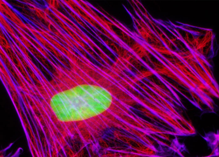
Male Rat Kangaroo Kidney Epithelial Cells (PtK2 Line)
In order to label the intermediate filaments in the PtK2 culture presented above, the fixed and permeabilized cells were blocked and treated with chicken anti-vimentin primary antibodies followed by goat anti-chicken secondary antibodies (IgG) conjugated to Alexa Fluor 568. Filamentous actin was visualized with phalloidin conjugated to Alexa Fluor 633, while the nuclei were counterstained with SYTOX Green. Images were recorded in grayscale with a 12-bit digital camera coupled to a Nikon Eclipse 80i microscope equipped with bandpass emission fluorescence filter optical blocks. During the processing stage, individual image channels were pseudocolored with RGB values corresponding to each of the fluorophore emission spectral profiles, with the exception of Alexa Fluor 633, which was pseudocolored magenta.
Featured in:
Share this page:


