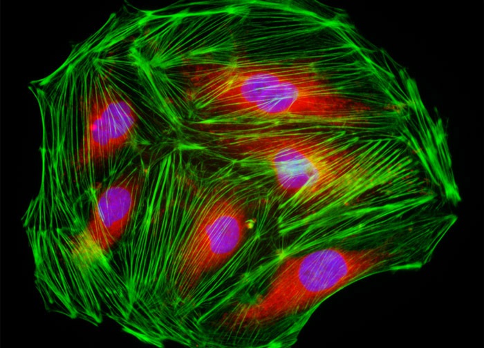
Male Rat Kangaroo Kidney Epithelial Cells (PtK2 Line)
The culture of rat kangaroo kidney epithelial (PtK2) cells featured in the digital image above was labeled with the enzyme DNase I conjugated to the fluorochrome Texas Red in order to reveal unpolymerized globular (G) actin, which accumulates in the center of the cell. In addition, the specimen was stained for DNA with the ultraviolet-absorbing probe DAPI, and for the cytoskeletal filamentous actin network with Alexa Fluor 488 conjugated to phalloidin. Images were recorded in grayscale with a 12-bit digital camera coupled to either a Nikon E-600 or Eclipse 80i microscope equipped with bandpass emission fluorescence filter optical blocks. During the processing stage, individual image channels were pseudocolored with RGB values corresponding to each of the fluorophore emission spectral profiles.
Featured in:
Share this page:


