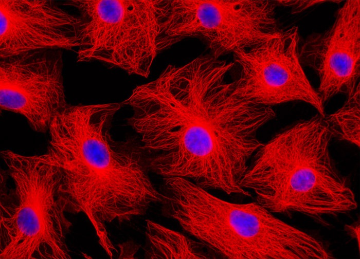
Male Rat Kangaroo Kidney Epithelial Cells (PtK2 Line)
In order to visualize the microtubules present in the rat kangaroo kidney cells illustrated in the digital image above, a PtK2 culture was immunofluorescently labeled with anti-tubulin mouse monoclonal primary antibodies followed by goat anti-mouse secondary antibody fragments conjugated to Rhodamine Red-X. In addition, the cells were labeled for cell nuclei with the classic nucleic acid stain, DAPI. Images were recorded in grayscale with a 12-bit digital camera coupled to either a Nikon E-600 or Eclipse 80i microscope equipped with bandpass emission fluorescence filter optical blocks. During the processing stage, individual image channels were pseudocolored with RGB values corresponding to each of the fluorophore emission spectral profiles.
Featured in:
Share this page:


