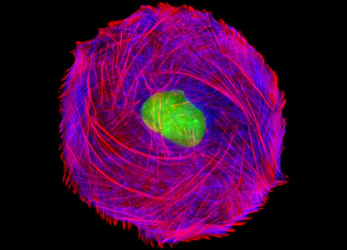
Mink Uterus Endometrium Epithelial Cells (GMMe Line)
The GMMe cells illustrated above were immunofluorescently labeled with primary anti-vimentin (an intermediate filament protein) mouse monoclonal antibodies followed by goat anti-mouse Fab fragments conjugated to Marina Blue. Alexa Fluor 594 conjugated to phalloidin (cytoskeletal F-actin network) and SYTOX Green (nuclei) were also used to stain the epithelial cell culture. Images were recorded in grayscale with a 12-bit digital camera coupled to either a Nikon E-600 or Eclipse 80i microscope equipped with bandpass emission fluorescence filter optical blocks. During the processing stage, individual image channels were pseudocolored with RGB values corresponding to each of the fluorophore emission spectral profiles.
Featured in:
Share this page:


