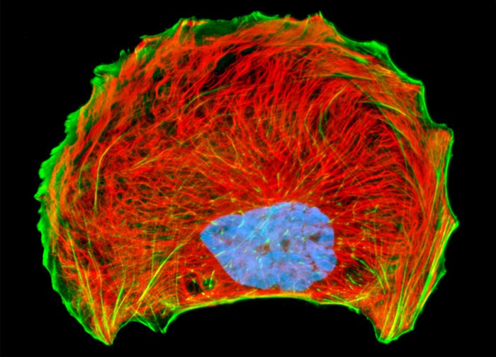
Mink Uterus Endometrium Epithelial Cells (GMMe Line)
The proximity of intermediate filaments and the cytoskeletal filamentous actin network was visualized by treating the fixed and permeabilized culture of mink uterus endometrium epithelial cells presented above with mouse anti-vimentin primary antibodies followed by goat anti-mouse secondary antibodies (IgG) conjugated to Texas Red-X. F-actin was subsequently labeled with Oregon Green 488 conjugated to phalloidin, and the nuclei were counterstained with Hoechst 33258. Images were recorded in grayscale with a 12-bit digital camera coupled to either a Nikon E-600 or Eclipse 80i microscope equipped with bandpass emission fluorescence filter optical blocks. During the processing stage, individual image channels were pseudocolored with RGB values corresponding to each of the fluorophore emission spectral profiles.
Featured in:
Share this page:


