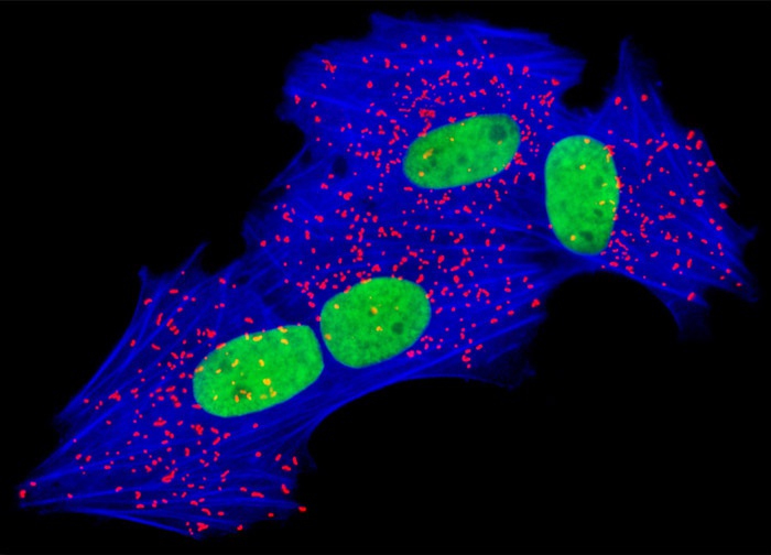
Mink Uterus Endometrium Fibroblast Cells (GMMs Line)
In a double immunofluorescence experiment, the adherent monolayer culture of mink endometrium fibroblast cells illustrated above was fixed, permeabilized, blocked with 10-percent normal goat serum, and then treated with a cocktail of mouse anti-histone and rabbit anti-PMP 70 (peroxisomal membrane protein 70) primary antibodies followed by goat anti-mouse and anti-rabbit secondary antibodies (IgG) conjugated to BODIPY FL and Alexa Fluor 568, respectively. The filamentous actin network was counterstained with Alexa Fluor 350 conjugated to phalloidin. Images were recorded in grayscale with a 12-bit digital camera coupled to either a Nikon E-600 or Eclipse 80i microscope equipped with bandpass emission fluorescence filter optical blocks. During the processing stage, individual image channels were pseudocolored with RGB values corresponding to each of the fluorophore emission spectral profiles.
Featured in:
Share this page:


