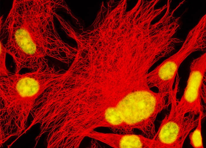
Mongolian Gerbil Lung Fibroblast Cells (GeLu Line)
Immunofluorescence with mouse anti-alpha-tubulin was employed to visualize distribution of the microtubule network in the gerbil lung fibroblast cell culture illustrated above. The secondary antibody (goat anti-mouse IgG) was conjugated to Alexa Fluor 568. DNA in the cell nucleus was labeled with the nucleic acid stain TO-PRO-3. Images were recorded in grayscale with a 12-bit digital camera coupled to either a Nikon E-600 or Eclipse 80i microscope equipped with bandpass emission fluorescence filter optical blocks. During the processing stage, individual image channels were pseudocolored with RGB values corresponding to each of the fluorophore emission spectral profiles with the exception of TO-PRO-3, which was pseudocolored yellow.
Featured in:
Share this page:


