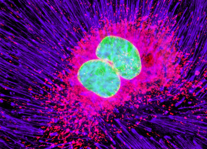
Mouse Hemangioendothelioma Endothelial Cells (EOMA Line)
The mouse hemangioendothelioma endothelial cells presented in the digital image above were resident in an adherent culture stained for F-actin with Alexa Fluor 633 conjugated to phalloidin and for mitochondria with MitoTracker Orange CMTMRos. DNA in the cell nucleus was counterstained with SYTOX Green. Images were recorded in grayscale with a 12-bit digital camera coupled to either a Nikon E-600 or Eclipse 80i microscope equipped with bandpass emission fluorescence filter optical blocks. During the processing stage, individual image channels were pseudocolored with RGB values corresponding to each of the fluorophore emission spectral profiles with the exception of Alexa Fluor 633, which was pseudocolored lavender.
Featured in:
Share this page:


