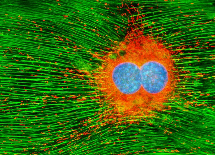
Mouse Hemangioendothelioma Endothelial Cells (EOMA Line)
The adherent monolayer EOMA cell culture presented in the digital image above was labeled for the cytoskeletal filamentous actin and intracellular mitochondrial networks with BODIPY FL conjugated to phalloidin (yielding green emission) and MitoTracker Orange CMTMRos, respectively. Nuclei present in the endothelial cells were counterstained with the DNA-selective bisbenzimide dye, Hoechst 33342 (blue emission). Images were recorded in grayscale with a 12-bit digital camera coupled to either a Nikon E-600 or Eclipse 80i microscope equipped with bandpass emission fluorescence filter optical blocks. During the processing stage, individual image channels were pseudocolored with RGB values corresponding to each of the fluorophore emission spectral profiles.
Featured in:
Share this page:


