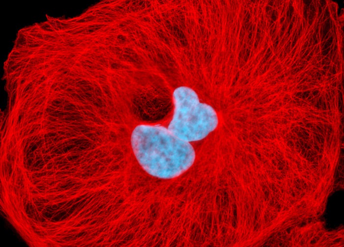
Normal African Green Monkey Kidney Fibroblast Cells (CV-1 Line)
The microtubules present in the log phase culture of CV-1 cells featured in the digital image presented above were immunofluorescently labeled with primary anti-tubulin mouse monoclonal antibodies followed by goat anti-mouse Fab fragments conjugated to Rhodamine Red-X. In addition, the culture was stained with Hoechst 33258, which selectively binds to DNA in cell nuclei. Images were recorded in grayscale with a 12-bit digital camera coupled to either a Nikon E-600 or Eclipse 80i microscope equipped with bandpass emission fluorescence filter optical blocks. During the processing stage, individual image channels were pseudocolored with RGB values corresponding to each of the fluorophore emission spectral profiles.
Featured in:
Share this page:


