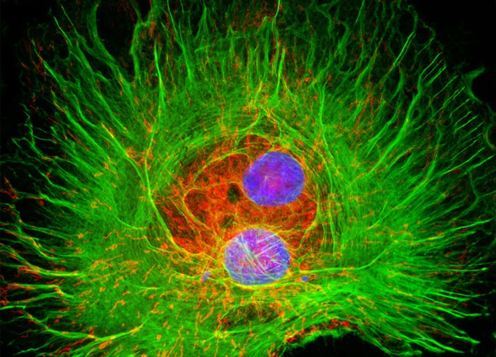
Normal African Green Monkey Kidney Fibroblast Cells (CV-1 Line)
The intracellular relationship between the cytoskeletal filamentous actin network and mitochondria present in a culture of CV-1 fibroblast cells (illustrated above) was visualized with the use of the probes Alexa Fluor 488 conjugated to phalloidin (yielding green fluorescence emission) and MitoTracker Red CMXRos. Cell nuclei were counterstained with DAPI (blue emission). Images were recorded in grayscale with a 12-bit digital camera coupled to either a Nikon E-600 or Eclipse 80i microscope equipped with bandpass emission fluorescence filter optical blocks. During the processing stage, individual image channels were pseudocolored with RGB values corresponding to each of the fluorophore emission spectral profiles.
Featured in:
Share this page:


