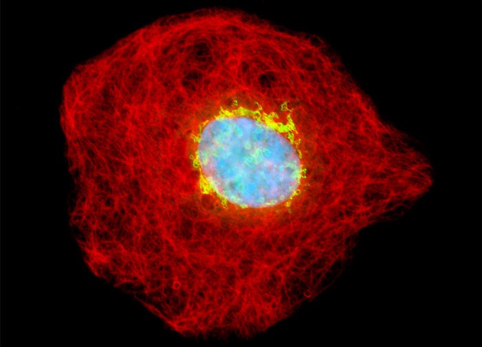
Owl Monkey Kidney Epithelial Cells (OMK Line)
The owl monkey kidney cells illustrated in the digital image above were fixed with paraformaldehyde, permeabilized, and treated with a mixture of rabbit (anti-giantin; Golgi complex) and mouse (anti-vimentin; intermediate filaments) primary antibodies, followed by secondary antibodies conjugated to Cy2 and Alexa Fluor 568, respectively. Cell nuclei were counterstained with the DNA-selective bisbenzimide dye, Hoechst 33258. Images were recorded in grayscale with a 12-bit digital camera coupled to either a Nikon E-600 or Eclipse 80i microscope equipped with bandpass emission fluorescence filter optical blocks. During the processing stage, individual image channels were pseudocolored with RGB values corresponding to each of the fluorophore emission spectral profiles.
Featured in:
Share this page:


