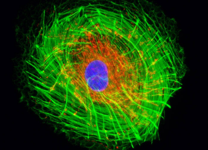
Rat Jejunum Myenteric Plexus Enteroglial Cells (EGC/PK060399egfr Line)
The mitochondrial and cytoskeletal F-actin networks were visualized in a culture of rat jejunum myenteric plexus enteroglial cells (illustrated above) by labeling them with MitoTracker Red CMXRos and Alexa Fluor 488 conjugated to phalloidin, respectively. Cell nuclei were counterstained with DAPI. Images were recorded in grayscale with a 12-bit digital camera coupled to either a Nikon E-600 or Eclipse 80i microscope equipped with bandpass emission fluorescence filter optical blocks. During the processing stage, individual image channels were pseudocolored with RGB values corresponding to each of the fluorophore emission spectral profiles.
Featured in:
Share this page:


