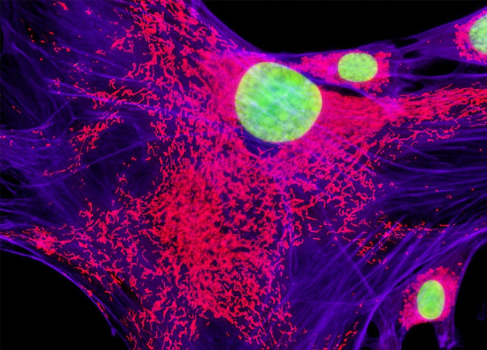
Swiss Mouse Embryo Moloney Murine Leukemia Virus Transfected Fibroblast Cells (CRE BAG 2 Line)
The cell culture featured in the digital image presented above was fluorescently labeled with MitoTracker Red CMXRos and SYTOX Green, targeting the mitochondrial network and cell nuclei, respectively. In addition, Alexa Fluor 633 conjugated to phalloidin was utilized to label filamentous actin. Images were recorded in grayscale with a 12-bit digital camera coupled to either a Nikon E-600 or Eclipse 80i microscope equipped with bandpass emission fluorescence filter optical blocks. During the processing stage, individual image channels were pseudocolored with RGB values corresponding to each of the fluorophore emission spectral profiles with the exception of Alexa Fluor 633, which was pseudocolored blue.
Featured in:
Share this page:


