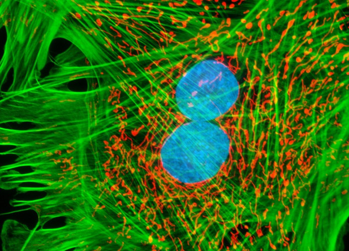
Tahr Ovary Epithelial Cells (HJ1.Ov Line)
The HJ1.Ov epithelial cells that appear in the digital image above were resident in a cell culture that was stained with MitoTracker Red CMXRos, Alexa Fluor 488 conjugated to phalloidin, and Hoechst 33342, which target the mitochondrial network, filamentous actin, and DNA in the cell nucleus, respectively. Images were recorded in grayscale with a 12-bit digital camera coupled to either a Nikon E-600 or Eclipse 80i microscope equipped with bandpass emission fluorescence filter optical blocks. During the processing stage, individual image channels were pseudocolored with RGB values corresponding to each of the fluorophore emission spectral profiles.
Featured in:
Share this page:


