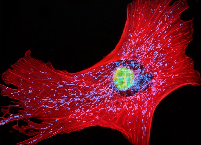
Tahr Ovary Epithelial Cells (HJ1.Ov Line)
The culture of tahr ovary epithelial cells presented in the digital image above was treated with MitoTracker Deep Red 633 and Alexa Fluor 568 conjugated to phalloidin, fluorescently labeling the mitochondrial network and F-actin, respectively. The nucleic acid stain YO-PRO-1 was utilized to counterstain cell nuclei. Images were recorded in grayscale with a 12-bit digital camera coupled to either a Nikon E-600 or Eclipse 80i microscope equipped with bandpass emission fluorescence filter optical blocks. During the processing stage, individual image channels were pseudocolored with RGB values corresponding to each of the fluorophore emission spectral profiles with the exception of MitoTracker Deep Red 633, which was pseudocolored cyan.
Featured in:
Share this page:


