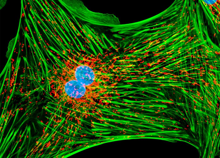
Tahr Ovary Epithelial Cells (HJ1.Ov Line)
The log phase monolayer culture of tahr ovary cells depicted above was treated with MitoTracker Red CMXRos in growth medium for one hour, washed, and fixed with 3.7-percent paraformaldehyde in medium containing serum. After washing and permeabilization, the cells were blocked with bovine serum albumen in PBS and labeled with BODIPY FL conjugated to phallacidin. The nuclei were subsequently counterstained with Hoechst 33258. Images were recorded in grayscale with a 12-bit digital camera coupled to either a Nikon E-600 or Eclipse 80i microscope equipped with bandpass emission fluorescence filter optical blocks. During the processing stage, individual image channels were pseudocolored with RGB values corresponding to each of the fluorophore emission spectral profiles.
Featured in:
Share this page:


