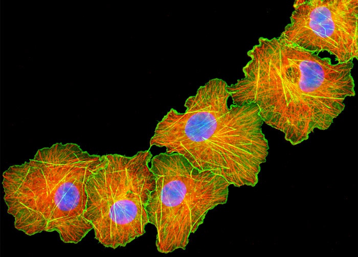
Transformed (Simian Virus 40) African Green Monkey Kidney Fibroblast Cells (COS-7 Line)
The microtubule network was visualized in the culture of transformed African green monkey kidney fibroblast cells (COS-7) displayed in the digital image above via immunofluorescent labeling with primary anti-tubulin mouse monoclonal antibodies followed by goat anti-mouse Fab fragments conjugated to Cy3. In addition, F-actin and cell nuclei were targeted with Alexa Fluor 488 conjugated to phalloidin and DAPI, respectively. Images were recorded in grayscale with a 12-bit digital camera coupled to either a Nikon E-600 or Eclipse 80i microscope equipped with bandpass emission fluorescence filter optical blocks. During the processing stage, individual image channels were pseudocolored with RGB values corresponding to each of the fluorophore emission spectral profiles.
Featured in:
Share this page:


