Laser Scanning Confocal Microscopy Video Gallery
The application of fluorescent proteins as intracellular probes for dynamics, localization, and gene expression has revolutionized the field of cell biology. The digital videos presented in this gallery explore a wide variety of these ubiquitous probes in fusions with subcellular localization peptides and proteins. Numerous cell lines expressing fluorescent protein fusions are imaged with a Nikon C1si microscope. Videos are presented in a streaming format, but they can also be downloaded as MPEG files or in a buffered progressive download format for repeated viewing.
Mitosis in Pig Kidney Epithelial Cells
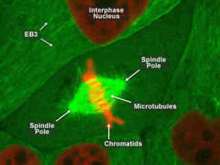
Examining mitosis in mammalian cell lines can reveal a number of details concerning the various activities associated with cell division. The digital videos in this section feature mitosis activities in a normal pig kidney (LLC-PK1) epithelial cell line stably expressing mCherry fluorescent protein fused to histone H2B and mEmerald fluorescent protein fused to human alpha-tubulin in live-cell culture.
EGFP and mCherry as Probes for Cell Division
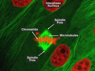
EGFP and mCherry as Probes for Cell Division - Substituting EGFP for the mEmerald fluorescent protein used to label alpha-tubulin in the previous section, the digital videos presented in this segment also examine mitosis events in normal pig kidney epithelial cells. mEmerald is a monomeric EGFP derivative that has been optimized with mutations designed to enhanced folding and maturation.
Investigating Actin and Mitochondria Dynamics
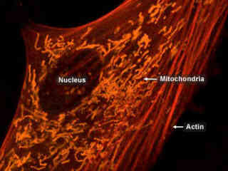
The videos presented in this section take advantage of spectral imaging and linear unmixing in laser scanning confocal microscopy to differentiate between actin and mitochondria labeled with overlapping orange and red fluorescent proteins. Normal Gray fox lung fibroblast cells were transfected with mCherry fluorescent protein fused to human beta-actin and a mitochondria targeting peptide fused to monomeric Kusabira Orange (mKO) fluorescent protein.
Golgi and Mitochondria Dynamics in Fibroblast Cells
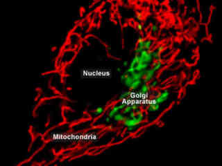
In order to visualize simultaneously the Golgi complex and mitochondria (and observe any potential interactions between these two organelles), fibroblast cells were transfected with a cocktail of EGFP fused to a Golgi targeting signal peptide and DsRed fluorescent protein fused to a mitochondrial targeting sequence. Imaging was conducted using argon-ion (488 nanometer) and diode-pumped solid state (561 nanometer) lasers.
Probing the Endoplasmic Reticulum with Green Fluorescent Proteins
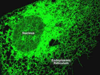
Proteins that assist in the folding/unfolding and the assembly/disassembly of other macromolecular structures are called chaperones. Within the endoplasmic reticulum (ER) there are many different families of chaperones with each family acting to aid protein folding in a different way.
Share this gallery:


