Ultraviolet Excitation: UV-2A (Longpass Emission)
The Nikon UV-2A ultraviolet fluorescence filter combination includes a medium-width bandpass excitation filter (330-380 nanometers) coupled to a longpass barrier filter (420 nanometer cut-on wavelength) to enable collection of signals from a wide spectrum of fluorescent probes. Ultraviolet and visible transmission spectral profiles for this filter combination are illustrated below in Figure 1. The common dichromatic mirror for the Nikon UV-2 series of filter combinations has a cut-on wavelength of 400 nanometers and reflects greater than 95 percent of the ultraviolet wavelengths between 330 and 390 nanometers.
Figure 1 - UV-2A (Standard Wide Band Ultraviolet Excitation)
Ultraviolet Excitation Filter Block UV-2A Specifications
- Excitation Filter Wavelengths: 330-380 nanometers (bandpass, 355 CWL)
- Dichromatic Mirror Cut-on Wavelength: 400 nanometers (longpass, LP)
- Barrier Filter Wavelengths: 420 nanometer cut-on (longpass, LP
The UV-2A fluorescence filter combination is designed to provide all-purpose imaging of fluorescent specimens with ultraviolet excitation, and is considered the standard set for this spectral region. The 50-nanometer bandpass excitation filter covers the longer ultraviolet wavelengths and is capable of exciting many of the fluorophores that have marginal absorption in this region. The longpass emission (barrier) filter employed in the UV-2Acombination is designed to collect signals at wavelengths exceeding 420 nanometers, enabling visualization of red, green, and blue emission from fluorophores excited in the ultraviolet. The UV-2A filter combination is recommended when studying the following fluorophores: Alexa Fluor 350, 7-amino-4-methylcoumarin (AMC), and its acetic acid derivatives (AMCA-S and AMCA), anthroyl stearate, bisaminophenyloxadiazole (BAO 9), bisbenzamide, Calcein Blue, Cascade Blue, dansyl derivatives, 4'-6-diamidino-2-phenylindole (DAPI), dopamine, Fast Blue, FluoroGold, Hoechst 33258/33342, noradrenaline, Nuclear Yellow, pyrene, tetracycline, and True Blue. The images presented in Figure 2 demonstrate the performance of the UV-2A filter combination with a variety of ultraviolet absorbing fluorescence probes targeted at different intracellular locations.
Figure 2 - Nikon UV-SA Wide Band Ultraviolet Excitation Longpass Filter Set
Illustrated in Figure 2(a) is the fluorescence emission intensity from a thin section of mouse intestine stained with Alexa Fluor 350 wheat germ agglutinin, a blue fluorescent lectin that is specific to the mucus of goblet cells. The ultraviolet absorption maximum of Alexa Fluor 350 is 346 nanometers and the emission maximum occurs at 442 nanometers. In addition, the specimen was simultaneously stained with Alexa Fluor 568 phalloidin (filamentous actin) and SYTOX Green (nuclei). Note the presence of spectral bleed-through from the red and green fluorophores and the overall low image contrast.
Figure 2(b) illustrates a culture of Indian Muntjac deerskin fibroblasts stained with Hoechst 33258, which specifically binds to DNA and stains the nuclei. The ultraviolet absorption maximum of Hoechst 33258 is 346 nanometers and the emission maximum is centered at 460 nanometers. Figures 2(c) and 2(d) present a mouse kidney thin section and bovine pulmonary artery cultured cells, respectively, both having the nuclei stained with DAPI (excitation at 358 nanometers and emission at 461 nanometers). Fluorescence emission centered at 422 nanometers from LysoTracker Blue DND-22, an ultraviolet probe that targets lysosomes, is presented in Figure 2(e).
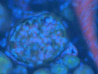
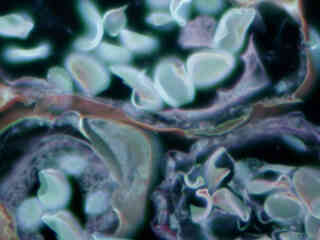
Autofluorescence in plant tissues (fern sporangium) is represented in Figure 2(f) and demonstrates the wide emission spectrum of endogenous fluorophores in these specimens. Note that images captured with the UV-2A filter combination have less contrast than images from the UV-2E/C set, but greater emission intensity than related combinations in the UV-1Aand UV-2B filter blocks.
Additional Specimen Images with the UV-2A Filter Combination
Mouse Intestine Thin Section
Fluorescence emission intensity from a thin section of mouse intestine stained with Alexa Fluor 350 wheat germ agglutinin, a blue fluorescent lectin that is specific to the mucus of goblet cells.
Indian Muntjac Nuclei
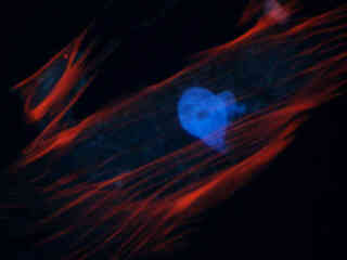
Fluorescence emission intensity from a culture of Indian Muntjac deerskin fibroblasts stained with Hoechst 33258, which specifically binds to DNA and stains the nuclei.
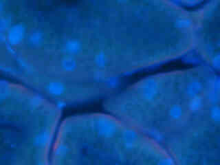
Mouse Kidney Tissue
Fluorescence emission intensity from a thin section of mouse kidney stained with multiple fluorophores.
Bovine Pulmonary Artery Cell Nuclei
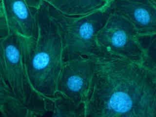
Fluorescence emission intensity from a culture of bovine pulmonary artery endothelial cells stained with DAPI, which targets DNA in the cell nucleus.
Contributing Authors
Related Nikon Products
Share this article:


