Are you on X?
Follow Nikon Instruments for microscopy news and trends, updates on new products, and information on contests and promotions.
October 30, 2019, via Nikon BioImaging Lab
We invite you to join us for the Nikon BioImaging Lab Opening Symposium & Luncheon, “Advanced Imaging Solutions for Neuroscience Drug Discovery” on Thursday, November 7 in Cambridge, MA! Space is limited, so please click to learn more and RSVP.
Learn More @ Nikon BioImaging Lab
October 21, 2019, via Nikon Small World

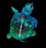
Nikon Instruments Inc. today announced the winners of the forty-fifth annual Nikon Small World Photomicrography Competition. First place was awarded to microscopy technician Teresa Zgoda and recent university graduate Teresa Kugler for their visually stunning and painstakingly prepared photo of a turtle embryo. Captured using fluorescence and stereo microscopy, the colorful final image is a masterful example of image-stitching.
Learn More @ Nikon Small World
January 18, 2019, via Nature
New research helps illuminate chemosensing in cancer cell invadopodia – protrusions of the cell membrane associated with increased metastasis. A Nikon A1R resonant scanning confocal was used to examine invadopodia dynamics in live embryos.
Learn More @ Nature
January 17, 2019, via Nature Communications Biology
Three new fluorescent protein-based biosensors for potassium! Both FRET-based and single-color versions are described, and simultaneous imaging of potassium and calcium dynamics is demonstrated.
Learn More @ Nature Communications Biology
January 15, 2019, via Nature Communications
Researchers identify the protein GORAB as an important coat protein scaffolding factor, helping to illuminate the mechanism behind the skin and bone disorder gerodermia osteodysplastica. Imaging for this study was performed in part using a Nikon Ti inverted microscope.
Learn More @ Nature Communications
January 14, 2019, via Johns Hopkins University
Johns Hopkins School of Medicine researchers demonstrate the importance of the signaling protein WISP-1 in regulating the differentiation of perivascular stem cells, helping our understanding of bone healing.
Learn More @ Johns Hopkins University
January 11, 2019, via Ars Technica
Scientists use induced pluripotent stem cells derived from autistic individuals as a model of neural development, identifying a group of genes expressing too early in the course of developing into the intermediate neural stem cell form.
Learn More @ Ars Technica
January 10, 2019, via Nature
Did you know that action potentials result in deformation of the cell membrane? Stanford researchers exploit this relation, using quantitative phase microscopy to capture action potentials by gentle all-optical imaging, and without fluorescent probes or electrodes.
Learn More @ Nature
January 07, 2019, via Science Immunology
New research shows that T cell antigen receptor engagement results in quick polymerization of nuclear actin into filaments needed to help drive effector functions. Video of nuclear actin polymerization upon immune synapse formation was performed on a Nikon Ti-E microscope with spinning disk confocal.
Learn More @ Science Immunology
January 04, 2019, via Nature Methods
Check out our app note in Nature Methods discussing the A1R HD25 confocal for live-cell imaging. The A1R HD25 features next-gen resonant scanner capable of covering the entire large 25mm FOV, great for live imaging of whole organisms such as the zebrafish embryo in the figure below.
Learn More @ Nature Methods
January 03, 2019, via Nature
Have you heard about the newly discovered graphene ‘magic angle’? Overlaying a pair of graphene sheets with their relative rotation set to the ‘magic angle’ results in unexpected superconductivity. There is also the possibility that this mechanism may be similar to those governing high-temperature superconductors.
Learn More @ Nature
December 28, 2018, via Cell Stem Cell
Researchers have succeeded in the direct reprogramming of human somatic cells into induced neural plate border stem cells (iNPBSCs) via ectopic expression of 4 factors. The iNPBSCs are self-renewing, multipotent, and easily modified with CRISPR/Cas9, with potential for applications in regenerative medicine.
Learn More @ Cell Stem Cell
December 26, 2018, via Science
Science Magazine chooses single cell RNA-seq for tracking gene expression in developing embryos as its “2018 Breakthrough of the Year”, noting the work of the Human Cell Atlas and others.
Learn More @ Science
December 21, 2018, via Science
The Boyden lab at MIT does it again with new Implosion Fabrication (ImpFab). Like Expansion Microscopy, ImpFab also exploits the shrinking/swelling properties of hydrogels to manipulate object size. ImpFab uses a hydrogel scaffold for arranging structures while at an easily workable size, followed by dehydration of the gel to shrink the entire assembly to a nano size. Importantly, this allows for new geometries that can’t be made through the typical process of assembly by stacking layers.
Learn More @ Science
December 19, 2018, via Nature Methods
New data from researchers at Kyoto University shows that cell membrane molecules are largely mobile following standard fixation protocols. The authors recommend fixation with 4% paraformaldehyde/0.2% glutaraldehyde for 30 min or longer.
Learn More @ Nature Methods
December 17, 2018, via Nature Communications
Open access research in Nature Communications details a new, higher sensitivity, tool for optogenetic silencing based on a photoactivated adenylyl cyclase and associated nucleotide-gated potassium channel. In vivo calcium imaging for this study was performed using a Nikon A1R MP resonant-scanning multiphoton system with gated NDDs.
Learn More @ Nature Communications
December 14, 2018, via Marine Biological Laboratory
Looking for a once-in-a-lifetime microscopy experience in Summer 2019? The Physiology Course at the Marine Biological Laboratory is looking for a new Microscopy Manager! Click for more details and application instructions.
Learn More @ Marine Biological Laboratory
December 13, 2018, via Nikon Instruments and Science
We have a new ebook published together with Science! Click to download your copy of Smarter imaging: Gaining more from your microscopy experiments.
Learn More @ Nikon Instruments and Science
December 06, 2018, via Nature
Did you know that bacteria can be forced to grow without a cell wall? New research shows that the formation of wall-less cells in actinomycetes may have evolved as an adaptation to osmotic stress. Imaging for this study was performed in part with a Nikon Ti inverted microscope with Yokogawa CSU-X1spinning disk confocal.
Learn More @ Nature
December 05, 2018, via Nature Communications
Alpha-catenin is a key protein linking the actin cytoskeleton with intercellular adhesions. Researchers at the University of Toronto shine new light on the mechanisms of tension-dependent F-actin binding by alpha-catenin.
Scratch wound assays for this study were performed using a Nikon IM-Q incubated microscope, and confocal imaging in part with a Nikon A1R resonant scanning system.
Learn More @ Nature Communications


