Nikon’s Museum of Microscopy
Model H Microscope
( early 1960s )
Resembling a classic Leica camera more than a microscope at first glance, the Nikon Model H field microscope was a laboratory-grade diagnostic instrument that found its way into the hands of physicians around the world. The circa-1960s Model H was often utilized in hemocytometry and in the detection of filariasis, malaria, and trypanosomiasis, also known as sleeping sickness.
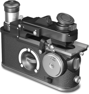
The Model H body is finished in chrome, black enamel, and leather, and like a fine camera, can be handheld, mounted on a tripod, or carried by a neck strap. A reinforced leather case safely stores the microscope, accessories, and an immersion oil dispenser, though it is not illustrated here. The Model H weighs about 800 grams and its design incorporates a high eyepoint, widefield, compensating eyepiece, a battery-powered illuminator, an Abbe condenser, and a fine focus wheel. For observation, specimens are mounted facedown under the clips on the crosswise traveling stage.
The optical path of the Model H, which is actually an inverted microscope, features a long-base prism typical of the circa-1930s John N. McArthur design for portable instruments. The objective changer knob provides the instrument's operator with a choice of three of the ten available parfocal, achromatic objectives that range in magnification from 4x to 100x. Six of the lenses are designed for brightfield/darkfield microscopy, while the other four are for phase contrast imaging. A special port on the side of the microscope allows the introduction of immersion oil when the 100x oil objective is engaged, and beneath the black body there is a diaphragm control and filter slot.
Share this page:


