Nikon’s Museum of Microscopy
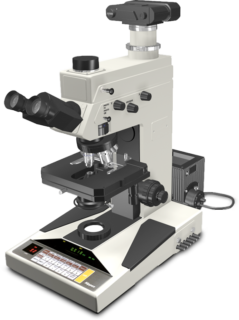
Microphot-FX Research Microscope
( Circa 1985 )
Promoted as one of the premier research microscopes during the mid-1980s, Nikon's Microphot-FX may be used with a digital or film camera attachment as well as for quantitative transmitted light and fluorescence microscopy. When utilized in combination with the Nikon Microflex FX camera series, it becomes a photomicrographic system and when coupled to a modern CCD camera, it becomes a digital imaging system.
Nikon, which is one of the few remaining optical companies that makes its own optical glass, designed an advanced optical system for the Microphot series with an emphasis on high performance in photomicrography. The lenses are made using an integrated production system, from the melting of the optical glass to the final assembly and adjustment. The goal is sharper, high contrast images with a minimum of chromatic, coma, astigmatism, and spherical aberration. Prominent characteristics of the objectives include a large numerical aperture for high resolving power, a greater flatness of the viewfield for more efficient observation, and improved parfocality, minimizing refocusing when objective magnification is changed.
Mechanical features of the Microphot were designed to increase potential applications and reduce operational complexity. The triaxial focus control provided three-speed control consisting of fine, medium, and coarse, in order to cover adjustments from ultra sensitive to long-range movement of the stage in a vertical motion. In addition to the rectangular mechanical stage, a circular polarizing stage was created for the Microphot. Nikon provided two eyepiece designs (interchangeable): one for the standard field width and one featuring ultra-widefield views (a field number of 26.5).
In order to cope with the demand for highly technical epi-fluorescence microscopy, a high-performance vertical illuminator attachment was prepared for the Microphot Series. The illuminator holds up to four interchangeable filter blocks, and turret rotation facilitates filter selection. A full line of 20 fluorescence filter blocks was available. The universal epi-illuminator system allowed the use of various reflected light examination techniques with a single nosepiece on one set of optics. Once the Epi-Illuminator is attached to the stand, a variety of microscopic observation modes, such as brightfield, darkfield, polarization, and DIC can be utilized in rapid succession, because no component reconfiguration is necessary to change examination techniques.
The Microphot phase contrast and differential interference contrast attachments are useful for observing the minute structures of unstained living cells or microorganisms. High-performance CF plan achromat and fluor phase objectives provide high-contrast phase images. The Bertrand lens built into the main body of the Microphot facilitates centering of the phase annular ring. The phase annular ring for each magnification is designed so that once centered in low magnification, no additional centering operation was needed for different magnifications.
Today Nikon manufactures a number of upright microscopes covering similar applications.
Share this page:


