Nikon’s Museum of Microscopy
Eclipse E1000
( 1997 )
The Nikon E1000 research-level microscope represented the top level in configurational options and performance of the Eclipse series of instruments, which were all equipped with the high-performance CFI60 optical system during the late 1990s. Advanced imaging capabilities (multiple camera ports) and several ergonomic features were designed to improve the quality of imaging and comfort of extended usage. With its introduction, the E1000 was perhaps one of the most sophisticated automatic research microscopes of the period.
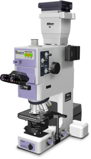
Ushering in a wholly different style of microscopy, the E1000 was the first automated microscope offering the look and feel of a standard microscope with the automation concealed from view. All necessary adjustments are instantly engaged upon changing from one objective to another, taking full advantage of motorized functions that provide optimum viewing. As many as seven micromotors are activated when the objective lens is changed, automatically setting the optimum aperture and field stop, brightness, condenser lens, and macroslider as well as configuration of other accessories included in the optical train. Automatic Kohler illumination and focusing (making automatic corrections of focal points) quickly establish ideal viewing conditions for many specimens. All these interconnected switching operations take place automatically every time the user changes objectives, with the fastest switching speed in the industry during the late 1990s.
The CFI60 specification is an optical system design that utilizes a wide array of Nikon technologies related to lens design, materials, and assembly techniques. Featuring high contrast and high resolution, it eliminates almost all flare, and offers high light transmission for fluorescence. The innovative objectives produced by Nikon enable long working distance with the capability for upgrading without sacrificing optical performance. Additionally, the CFI60 system ensures brightfield observation with high levels of resolution and contrast, minimal flare and a high transmission factor over the entire field of view. As an added feature and to minimize stress and provide an intuitive operating environment, the E1000 automatically makes the changes and adjustments necessary to optimize the quality of each image.
In the E1000, the revolving nosepiece and condenser are operated by a combination of mechanical and electrical controls to ensure accuracy and durability. A self-correcting mechanism compensates for errors likely to occur as the microscope ages. When rotating objectives between wet and dry slides, the nosepiece automatically stops halfway between the objectives, facilitating the addition of media to oil or water-immersion slides or to clean the objective front lens and slide when switching to a dry specimen.
An RS-232C interface is provided so the microscope can be controlled by a computer for telepathology, image analysis, or digital imaging applications. A unique bar code system reduces data entries by up to 80 percent. Specifications for each objective mounted on the nosepiece are recorded and the microscope can be configured by scanning a bar code list. Each of the corresponding settings, including optimum apertures and field stops, neutral density filter requirements and other settings necessary for automatic Kohler illumination are stored in the memory of the system.
Manual adjustments made to correct apertures and field stops, brightness and other items following the standard set-up can be stored on an IC smart card for instant recall when needed. This timesaving feature is convenient when viewing or comparing large volumes of specimens under identical conditions or in situations in which more than one researcher is using the E1000.
Featuring a modular design, the photo system head is integrated into the main microscope body, and offers the capability of advanced photomicrography in multiple formats. The E1000 optical system allows 100 percent of the light to enter all ports, providing ideal conditions for virtually every conceivable form of digital imaging. A digital camera, dual 35 millimeter film cameras, and a large-format 4 x 5 film camera can be mounted simultaneously.
To enhance contrast in transparent specimens, the de Sénarmont method of differential interference contrast (DIC) was adopted to optimize comfort and performance. By rotating the polarizer on the field lens, the DIC system is able to make precise adjustments to bias retardation to optimize contrast in the specimen under observation.
For more versatility when switching between phase contrast, fluorescence, brightfield and DIC observations, the E1000 offers Plan Fluor DLL objectives having high transmission phase rings in the rear focal plane. The phase contrast technique ensures sharp images with exceptional contrast and neutral background coloration, regardless of the magnification, and minimizes light loss when imaging with other modalities.
Several ergonomic features allow for operation of the E1000 in a natural position. The design of the E1000 enables the user to operate the focus knobs, stage handle and objective change buttons using one hand, freeing the other hand to perform supplementary functions. The knobs and stage handle are on the frame base at equal distances from the user and placed to permit a natural and comfortable hand and sitting position. The eyepiece tube is elevated to a viewing angle of 20 degrees, and an optional variable tilting eyepiece allows the user to set the eye level at the most natural and comfortable viewing position.
Today the Eclipse E1000 is succeeded by Nikon's fully motorized Eclipse Ni-E upright microscope for advanced research applications.
Share this page:


