Nikon’s Museum of Microscopy
Slit Lamp CS-1 Microscope
( Circa 1976 )
Nikon slit lamp microscopes are used by ophthalmologists to directly examine the eyes of a patient under the magnification of a binocular microscope by creating a stereoscopic, erect image. Known also as biomicroscopes, slit lamp microscopes, like the Nikon CS-1 illustrated below, are based on the 1902 design of Swedish ophthalmologist and Nobel Laureate, Allvar Gullstrand.
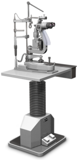
The slit lamp CS-1 microscope produces a narrow beam of intense light that can illuminate the patient's cornea, aqueous humor, crystalline lens, anterior vitreous layer, or other transparent ocular tissue. A form of oblique microscopy, the slit lamp microscope uses an imaging technique similar to optical sectioning. Interchangeable paired 10x and 15x eyepieces fit the binocular tubes of the CS-1 biomicroscope. Thus, the ophthalmologist controls the magnification during an eye examination. Each ocular lens features a diopter adjustment ring and a movable mount for matching an examiner's interpupillary distance and visual acuity. Objectives for the Nikon slit lamp microscope are relatively low-powered 1.0x and 1.6x lenses, which combine with the eyepieces for total magnifications ranging from 10x to 24x. The numerical aperture of 0.04 for the lower power objective and 0.07 for the 1.6x lens are notably very low to ensure maximum illumination and penetration into the eye tissues, with a more concentrated beam of light. A relatively long working distance of 101.5 millimeters is provided for the patient's comfort and to facilitate accurate diagnoses, even for the deeper eye layers.
A 6-volt, 30-watt tungsten-halogen bulb provides the light for the CS-1 slit lamp illumination system that features a brightness adjustment dial. The slit adjusts from 0.0 to 9 millimeters in width and ranges from 0.2 to 9 millimeters in length, depending on the setting of the five-position turret. Maximum rotation of the slit from the initial vertical position is 90 degrees in either direction via the slit rotating lever, which is mounted above the lamp housing. Green and cobalt blue filters are provided as accessories for the CS-1 illuminator and can be brought into the optical path by moving the filter change-over lever. The gelatin green filter blocks red light, which aids observation of the blood vessels in the sclera and conjunctiva membranes.
Employment of the blue filter, when coupled with an applanation-tonometer, facilitates the measurement of intraocular pressure and the diagnosis of glaucoma. To position the lamp, the focusing rod is erected and it is adjusted until the black surface faces the operator. For the fine motion of the slit image, a lever that resembles a video game joystick is tilted and for coarse motions, the lever is moved in the desired direction. The lamp housing is adjusted vertically and is centered until the image of the slit is projected onto the black surface and there is even illumination across the field of view.
The Nikon slit lamp CS-1 microscope illustrated above is mounted on a flat metal base plate for stability. The entire visual diagnostic apparatus is raised or lowered by two foot pedal switches mounted on a combination power control unit and telescoping instrument pedestal. The table adds stability and helps control unwanted vibrations during eye exams. For finer vertical adjustments to match the patient's height and eye position, the level adjustments for the instrument body and the chin rest are moved up or down, until the slit image is projected on the patient's eye correctly. Once the patient's head is set against the forehead and chin rest, the width of the slit is adjusted by turning the slit-width change knob that is mounted on top of the joystick lever, and which is located nearest the ophthalmologist on the microscope base. A fixation lamp enables the patient to focus the eye in a standard direction and distance, while a reference line on the post allows the doctor to examine the patient in a normal or standardized position. The Hruby lens, when placed in front of the patient's eye, allows the doctor to focus from the rear vitreous humor back into the fundus region of the eye.
Today Nikon is proud to distribute imaging products for ophthalmology under the Optos brand.
Share this page:


