The investigation of disease in humans has, understandably, been one of the primary focal points in medicine for thousands of years. The image gallery presented in this section attempts to illustrate, through use of the brightfield microscope, many of the pathological conditions that are readily observed in stained human specimens. Each image was chosen for artistic merit, photographic quality, and content. Note that several of the images in this gallery might not depict every aspect of the pathological condition under which they are catalogued.
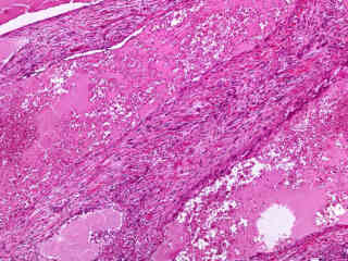
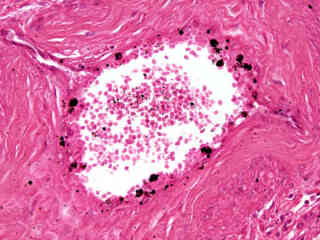
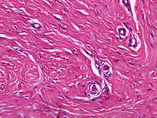
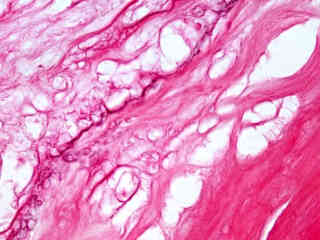
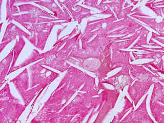
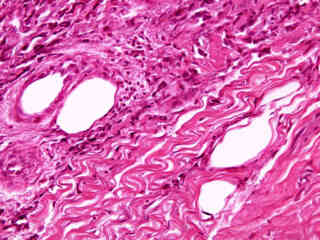
Aortic Atherosclerosis (Older Lesion)
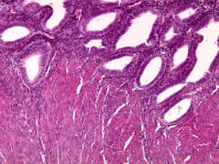
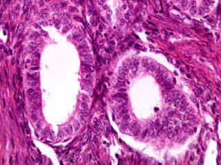
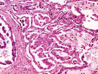
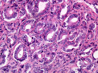
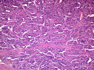
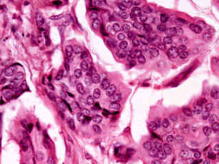
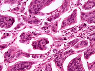
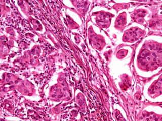
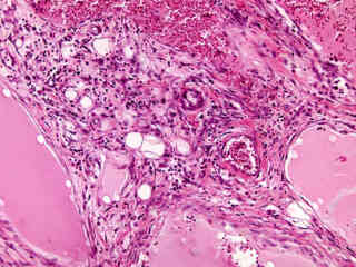
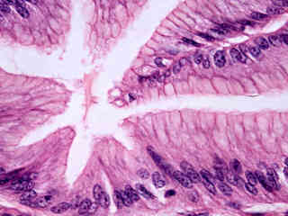
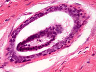
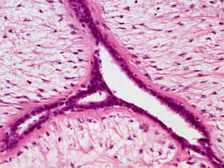
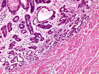
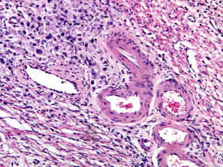
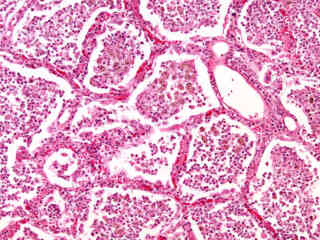
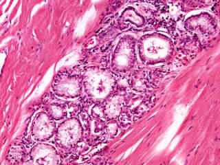
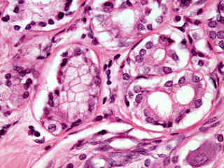
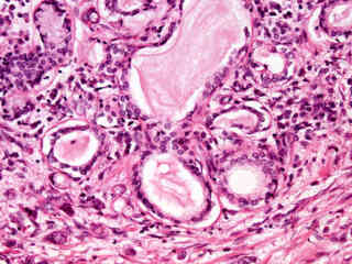
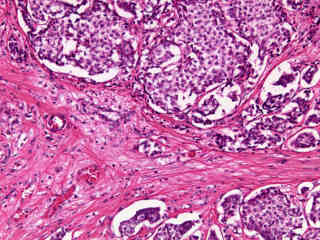
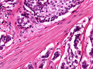
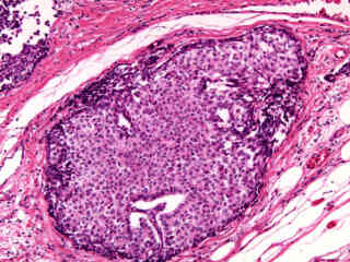
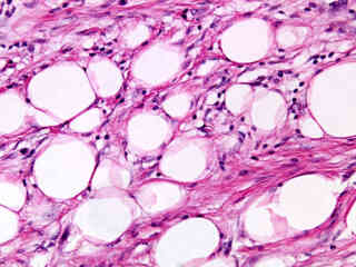
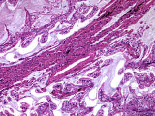
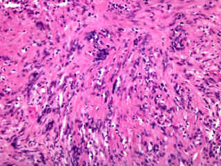
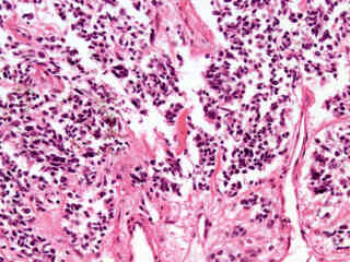
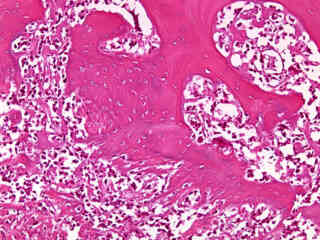
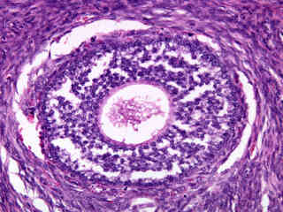
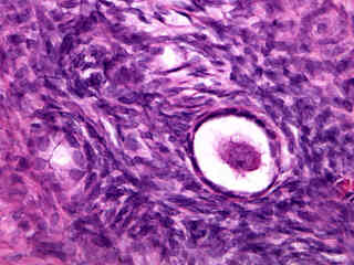
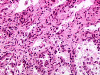
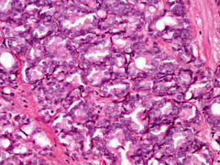
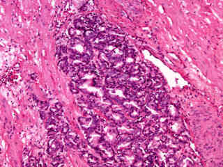
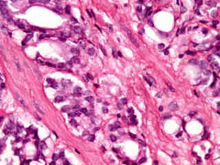
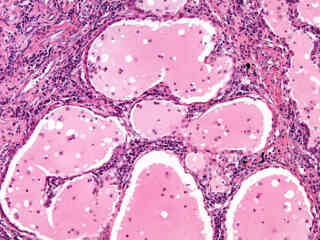
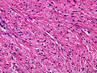
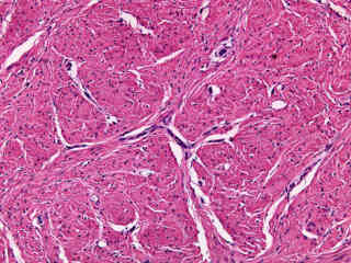
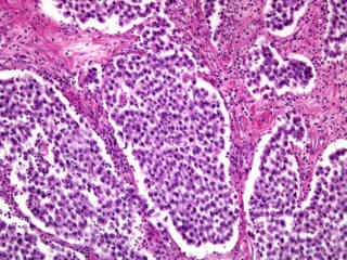
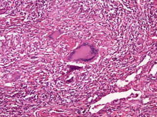
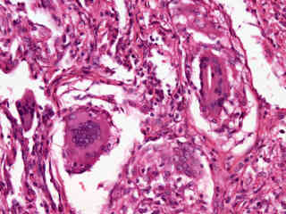
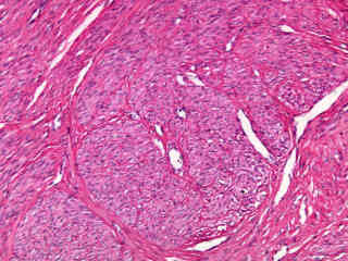
Benign Prostatic Hyperplasia
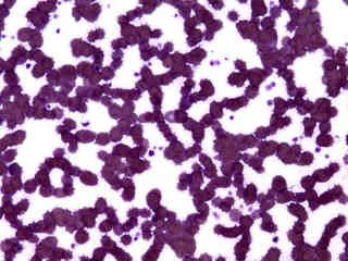
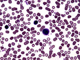
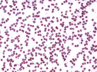
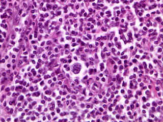
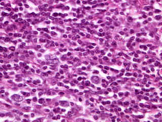
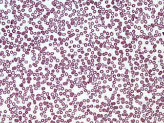
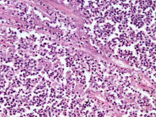
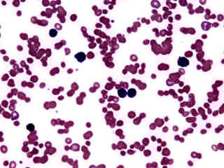
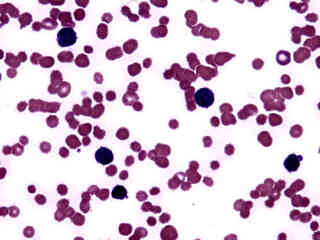
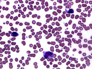
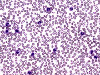
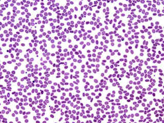
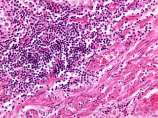
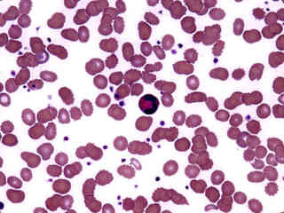
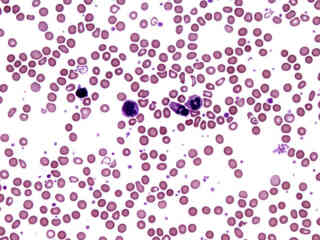
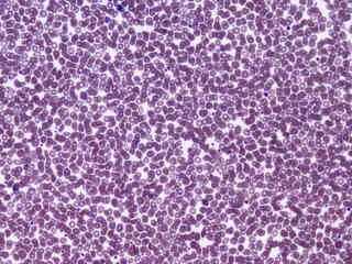
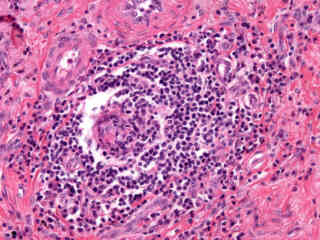
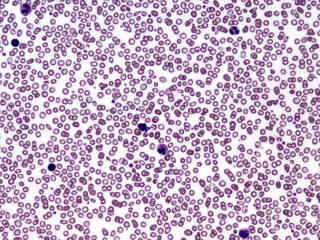
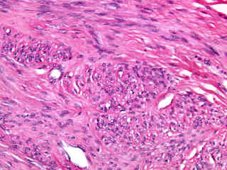
Chronic Lymphocytic Leukemia
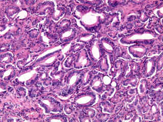
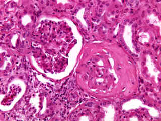
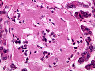
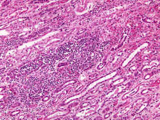
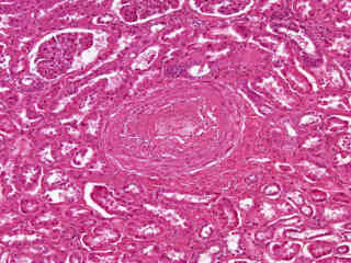
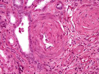
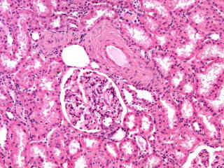
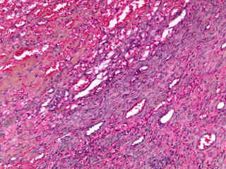
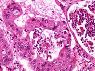
Diabetes in Kidney Tissue
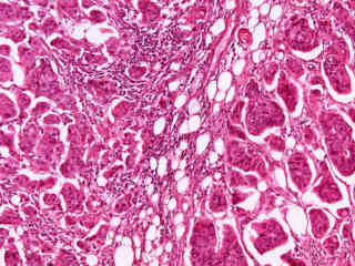
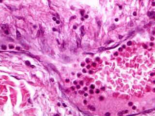
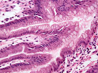
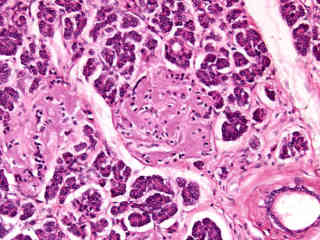
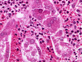
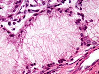
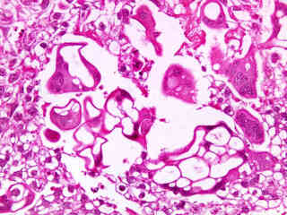
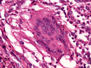
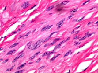
Diabetes Mellitus in Pancreatic Tissue
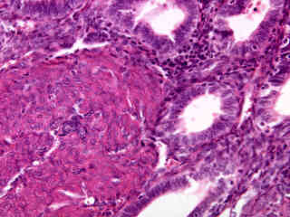
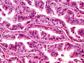
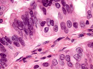
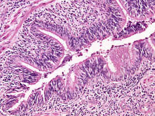
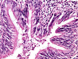
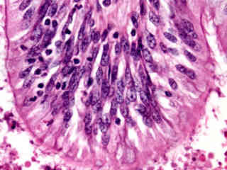
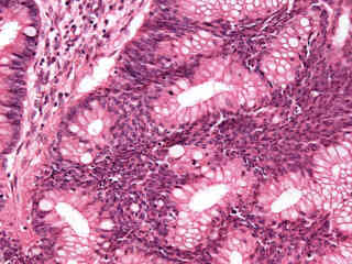
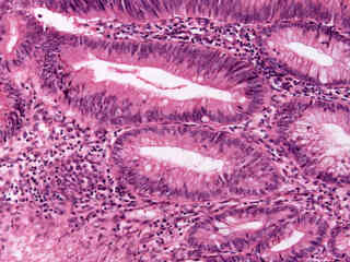
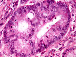
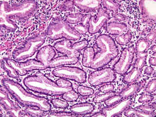
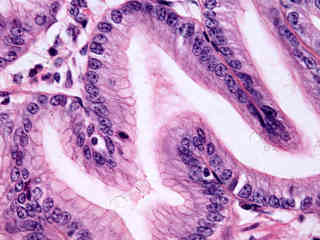
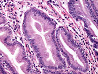
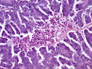
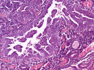
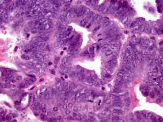
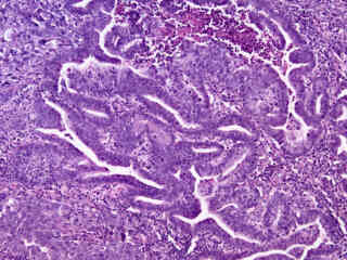
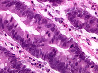
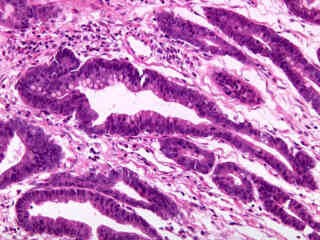
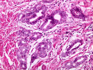
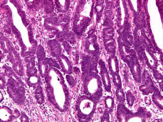
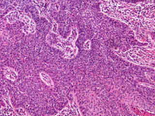
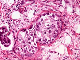
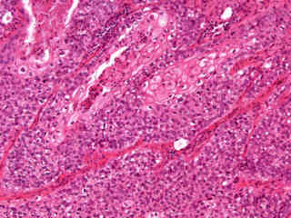
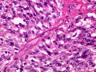
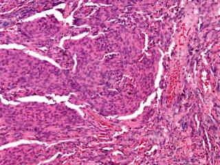
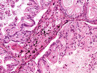
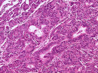
Endometrial Adenocarcinoma
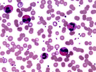
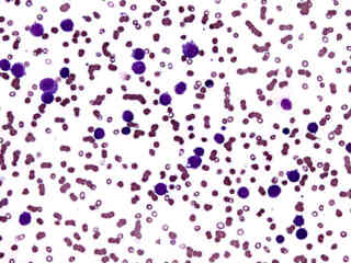
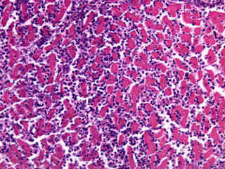
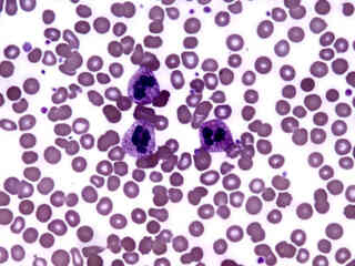
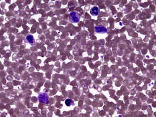
Granulocytic Leukemia (Acute)
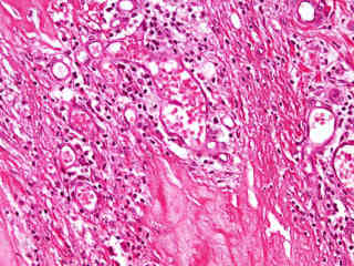
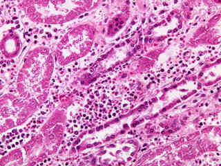
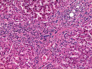
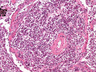
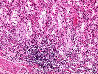
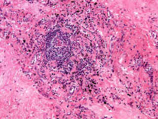
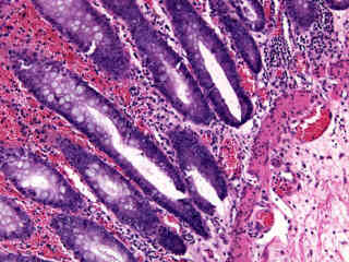
Hematogenous Pyelonephritis
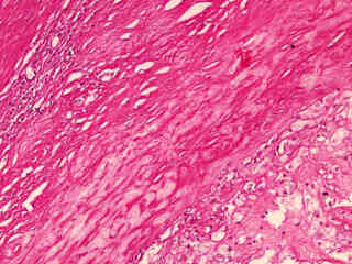
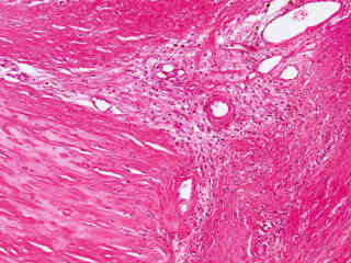
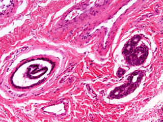
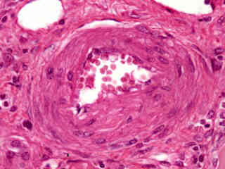
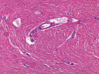
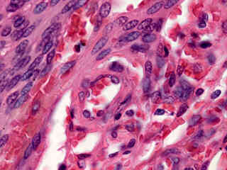
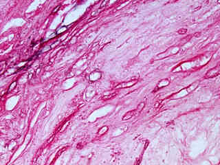
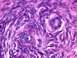
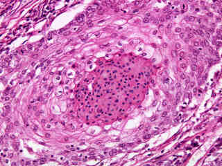
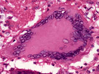
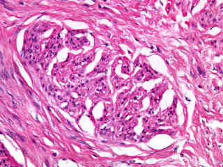
Hyperplastic Arteriosclerosis
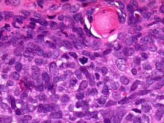
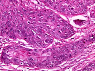
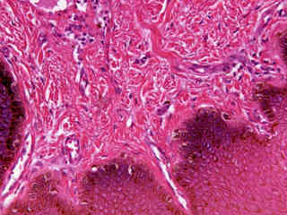
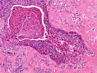
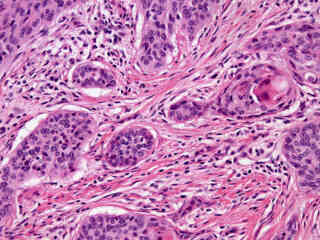
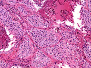
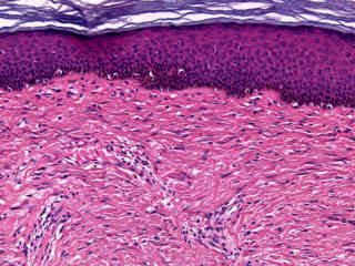
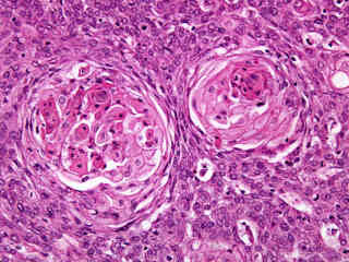
Laryngeal Squamous Cell Carcinoma
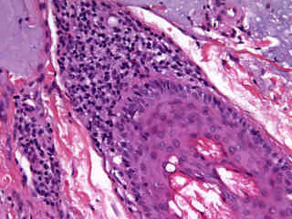
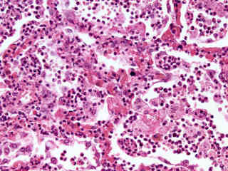
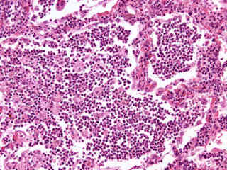
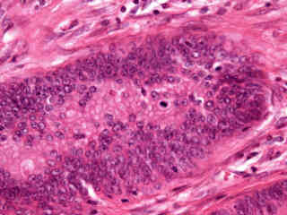
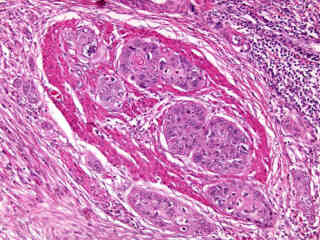
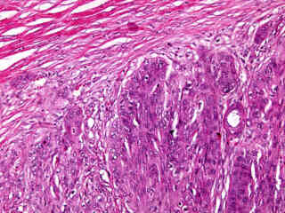
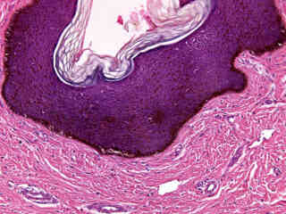
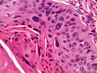
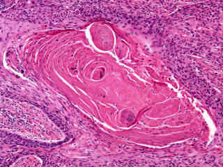
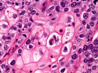
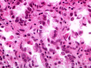
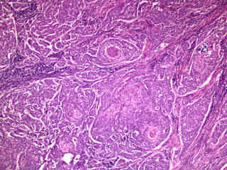
Lung Epidermoid Carcinoma
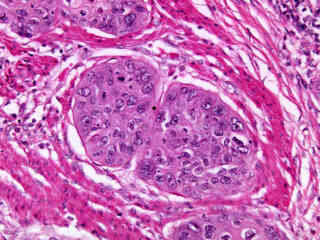
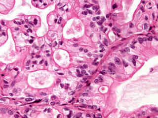
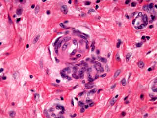
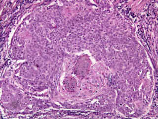
Lymph Node Metastatic Carcinoma
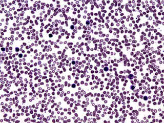
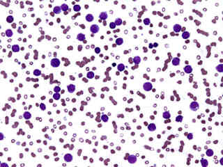
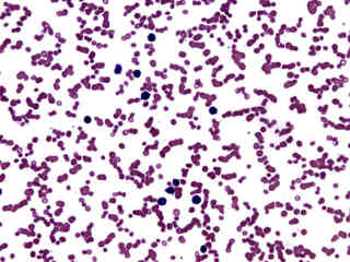
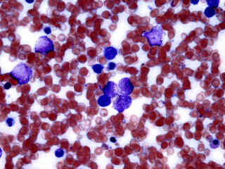
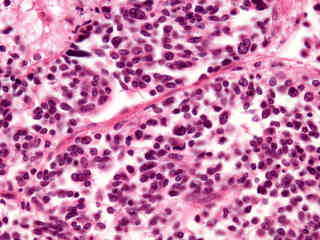
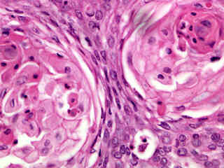
Lymphocytic Leukemia (Acute)
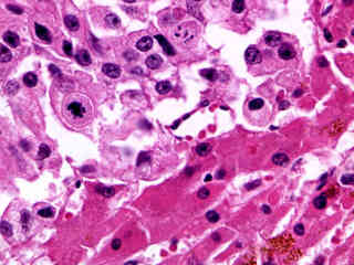
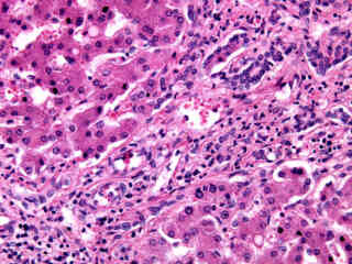
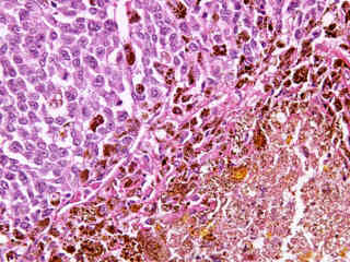
Metastatic Carcinoma in Liver Tissue
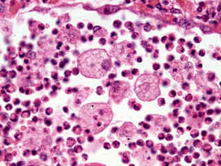
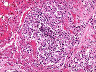
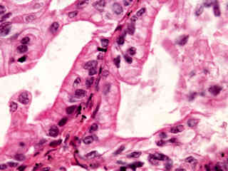
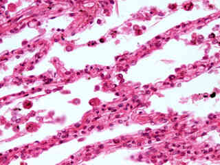
Metastatic Carcinoma in Lung Tissue
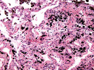
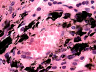
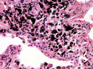
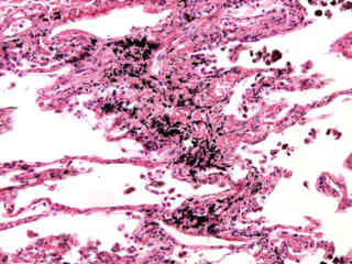
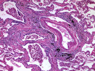
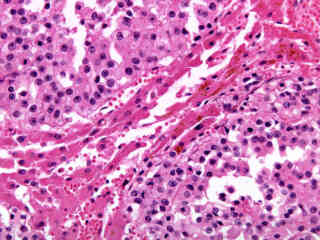
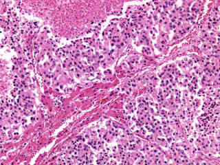
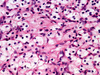
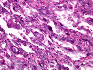
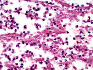
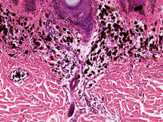
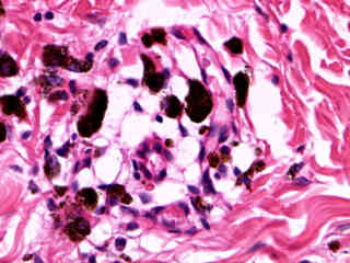
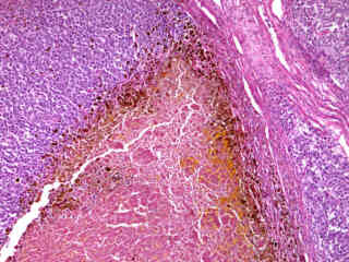
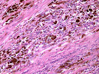
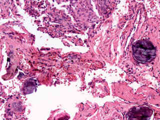
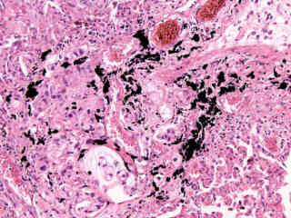
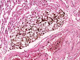
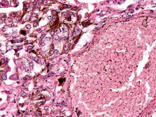
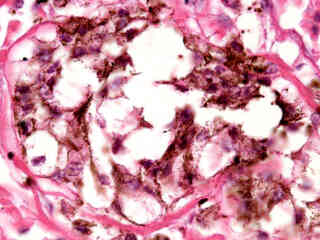
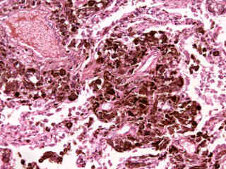
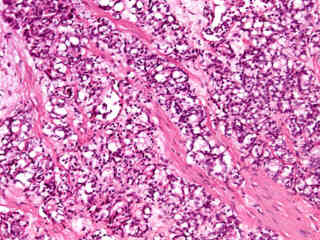
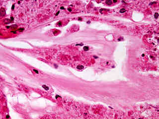
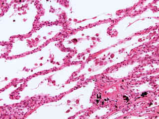
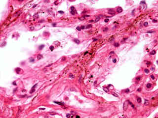
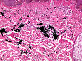
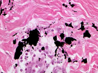
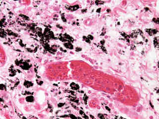
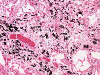
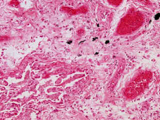
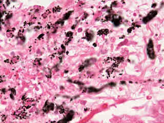
Metastatic Melanoma in Lung Tissue
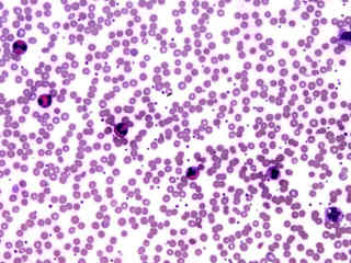
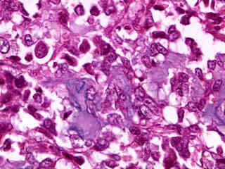
Myelomonocytic Leukemia (Acute)
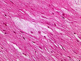
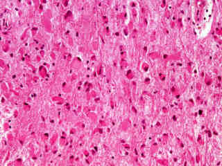
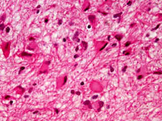
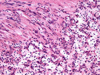
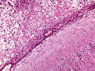
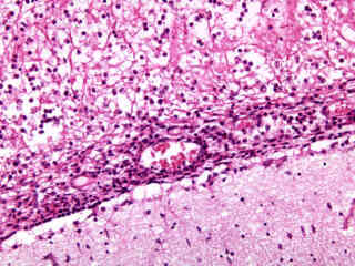
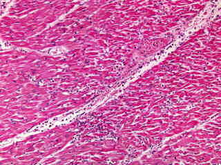
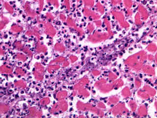
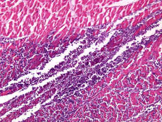
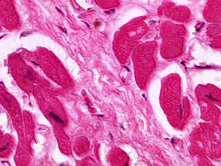
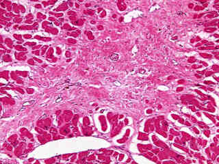
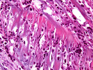
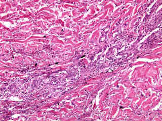
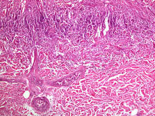
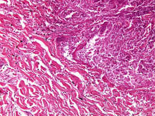
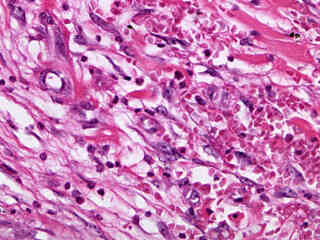
Myocardial Infarction (Acute)
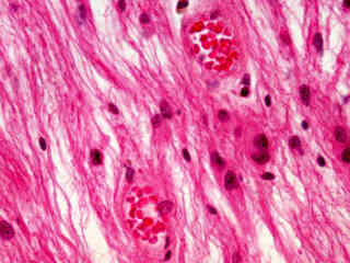
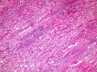
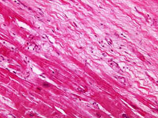
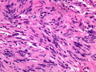
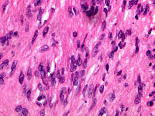
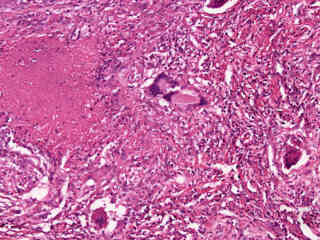
Myocardial Infarction (Old)
Occlusive Atherosclerosis
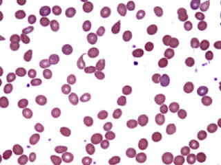
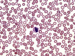
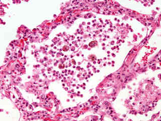
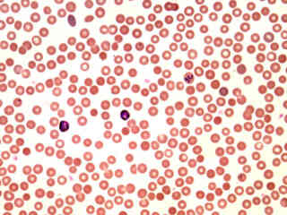
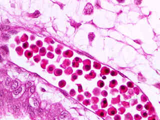
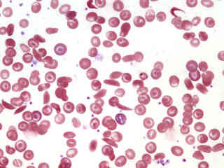
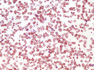
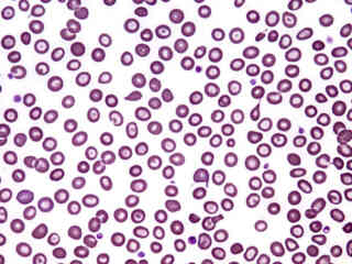
Plasmodium vivax Infection
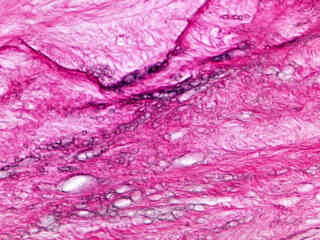
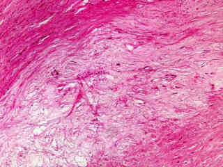
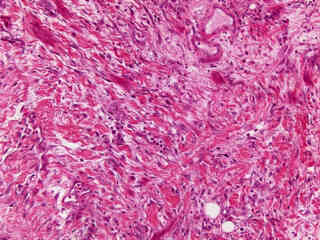
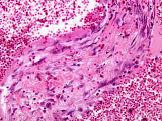
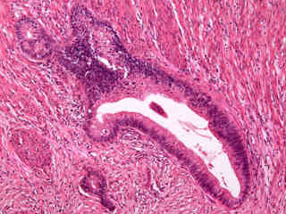
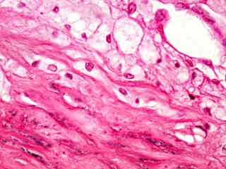
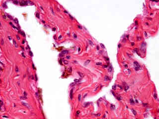
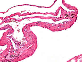
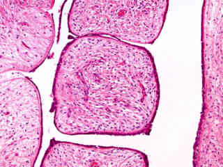
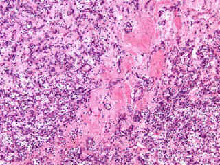
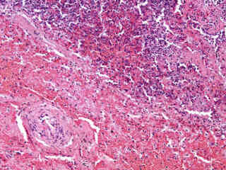
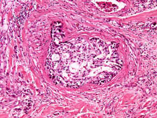
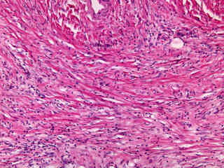
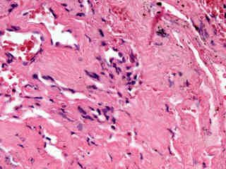
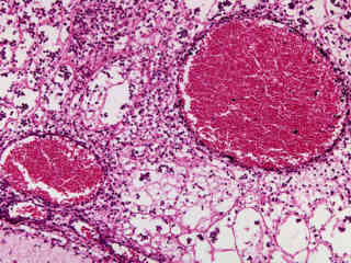
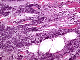
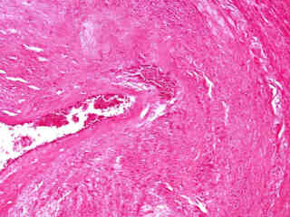
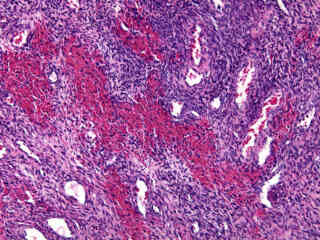
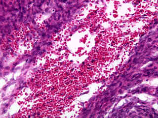
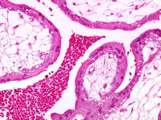
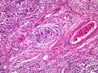
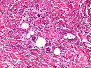
Ruptured Ectopic Pregnancy
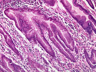
Suppurative Appendicitis (Acute)