The fluorescence microscope provides an interesting window into the world of the cell and is one of the biologist's favorite tools for the examination of both living and fixed cells in culture. The ability to specifically target organelles and macromolecules with synthetic fluorophores and immunofluorescence has produced a virtual revolution in the dynamic field of fluorescence microscopy for the examination of cells in culture. This portion of the fluorescence digital image gallery features widefield fluorescence images captured from over 30 cell lines stained with a mixture of synthetic probes, antibodies, and fluorescent proteins.
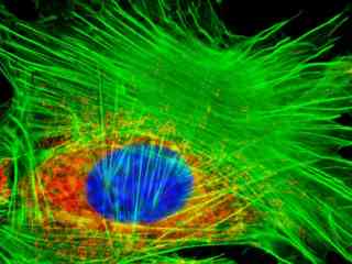
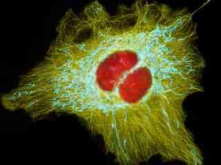
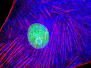
Normal African Green Monkey Kidney Fibroblast Cells (CV-1 Line)
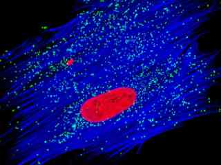
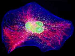
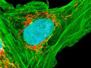
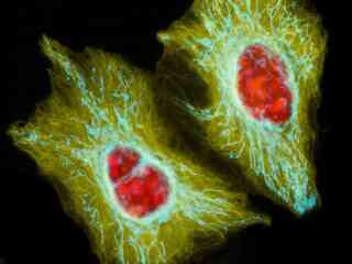
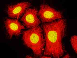
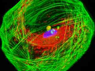
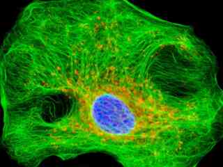
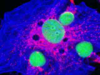
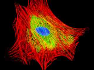
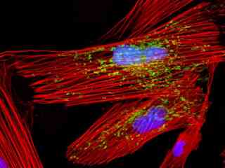
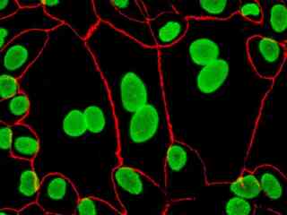
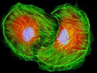
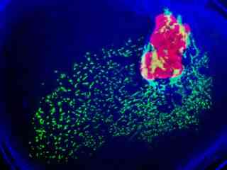
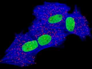
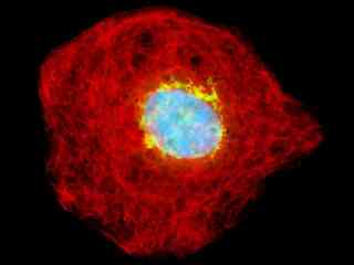
Transformed (Simian Virus 40) African Green Monkey Kidney Fibroblast Cells (COS-1 Line)
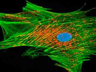
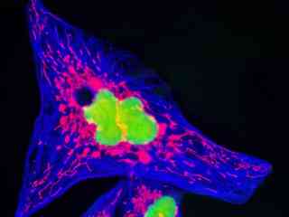
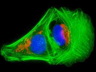
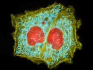
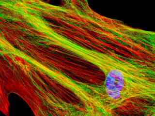
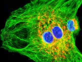
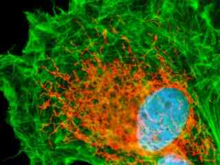
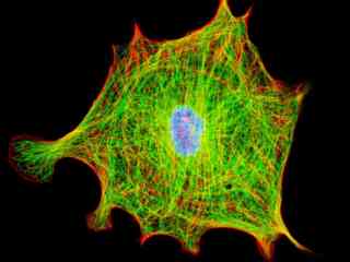
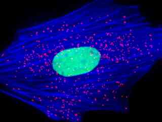
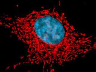
Transformed (Simian Virus 40) African Green Monkey Kidney Fibroblast Cells (COS-7 Line)
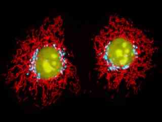
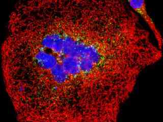
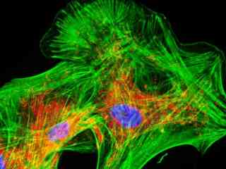
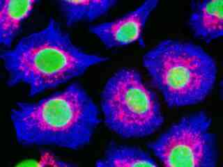
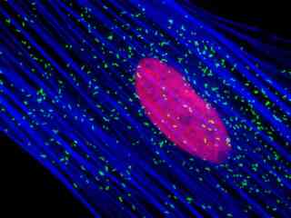
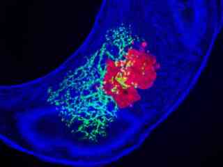
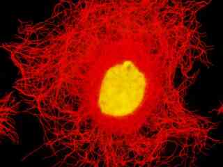
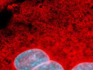
African Water Mongoose Skin Fibroblast Cells (A.P. Mongoose Line)
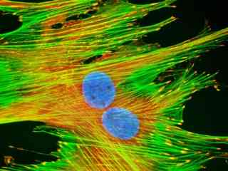
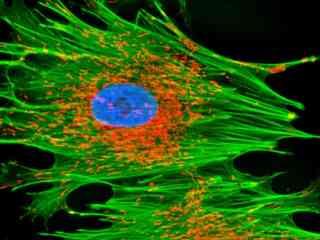
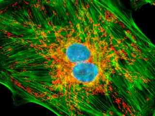
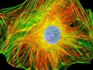
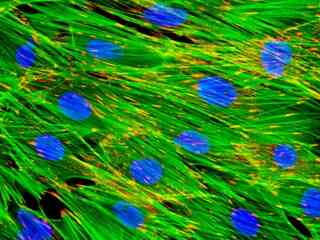
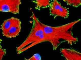
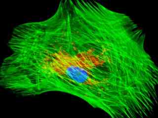
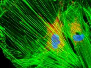
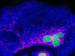
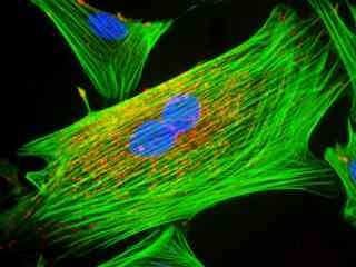
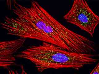
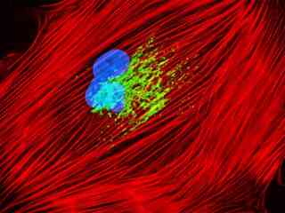
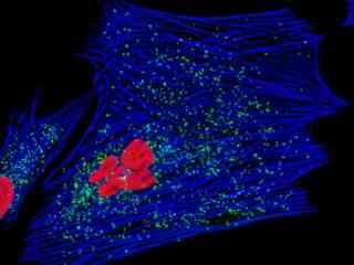
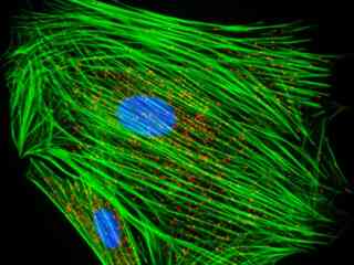
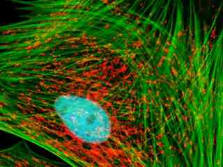
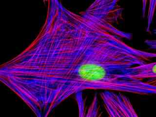
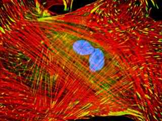
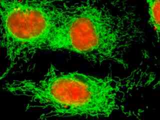
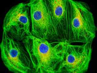
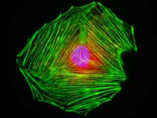
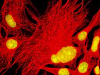
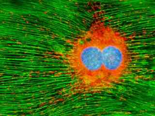
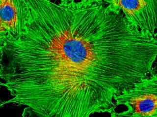
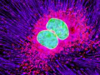
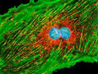
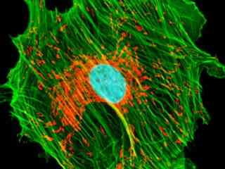
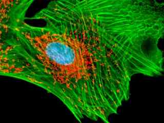
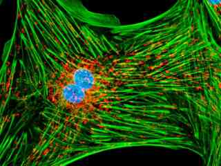
Bovine Pulmonary Artery Endothelial Cells (BPAE Line)
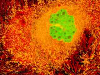
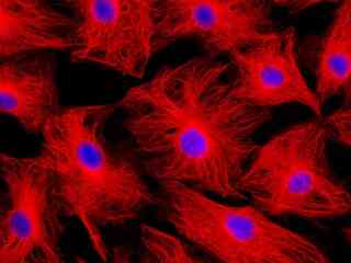
Chinese Hamster Ovary Cells (CHO-K1 Line)
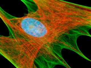
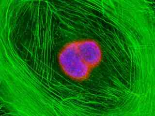
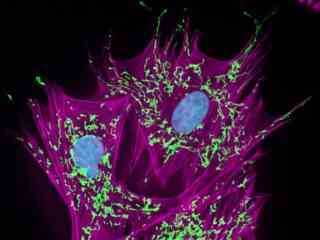
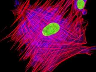
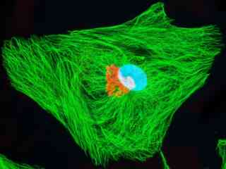
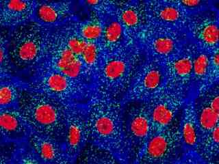
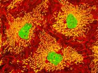
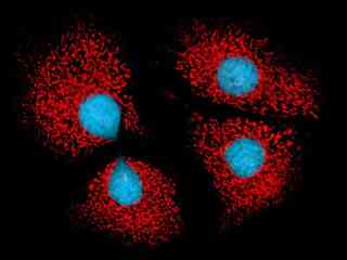
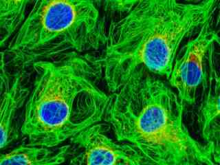
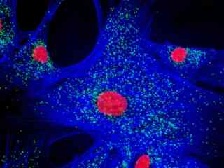
Embryonic Swiss Mouse Fibroblast Cells (3T3 Line)
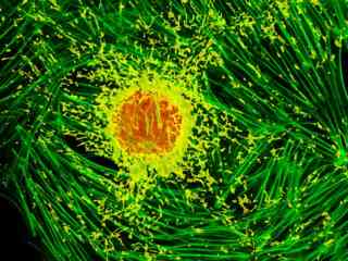
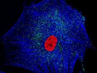
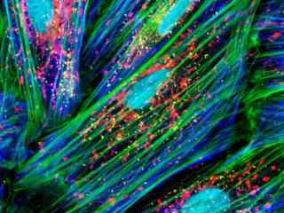
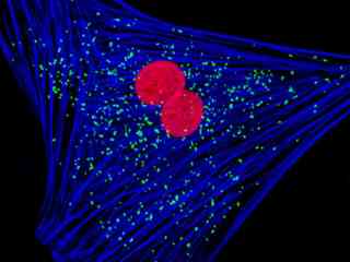
Embryonic Rat Thoracic Aorta Medial Layer Myoblast Cells (A-10 Line)
Embryonic Rat Thoracic Aorta Smooth Muscle Fibroblast Cells (A7r5 Line)
Grey Fox Lung Fibroblast Cells (FoLu Line)
Guinea Pig Colorectal Adenocarcinoma Epithelial Cells (GPC-16 Line)
Horse Dermal Fibroblast Cells (NBL-6 Line)
Human Bone Osteosarcoma Cells (U-2 OS Line)
Human Brain Glioma Cells (U-118 MG Line)
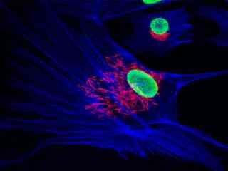
Human Cervical Adenocarcinoma Cells (HeLa Line)
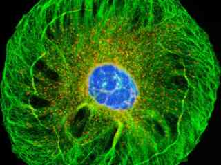
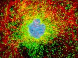
Human Cortical Neuronal Cells (HCN-1A Line)
Human Fetal Lung Fibroblast Cells (MRC-5 Line)
Human Lung Carcinoma Cells (A-549 Line)
Iguana Heart Epithelial Cells (IgH-2 Line)
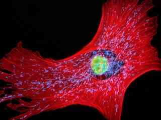
Indian Muntjac Deer Skin Fibroblast Cells
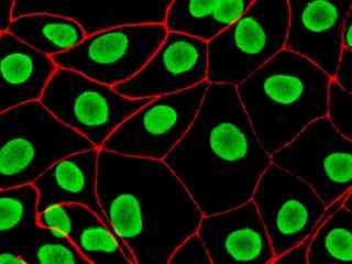
Madin-Darby Canine Kidney Epithelial Cells (MDCK Line)
Madin-Darby Ovine Kidney Epithelial Cells (MDOK Line)
Male Rat Kangaroo Kidney Epithelial Cells (PtK2 Line)
Mink Uterus Endometrium Epithelial Cells (GMMe Line)
Mink Uterus Endometrium Fibroblast Cells (GMMs Line)
Mongolian Gerbil Lung Fibroblast Cells (GeLu Line)
Mouse Hemangioendothelioma Endothelial Cells (EOMA Line)
Opossum Kidney Cortex Epithelial Cells (OK Line)
Owl Monkey Kidney Epithelial Cells (OMK Line)
Rabbit Kidney Epithelial Cells (RK13 Line)
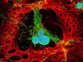
Rat Jejunum Myenteric Plexus Enteroglial Cells (EGC/PK060399egfr Line)
Rhesus Monkey Kidney Epithelial Cells (LLC-MK2 Line)
Swiss Mouse Embryo Moloney Murine Leukemia Virus Transfected Fibroblast Cells (CRE BAG 2 Line)
Tahr Ovary Epithelial Cells (HJ1.Ov Line)